Dynamics of aptamers
Leveraging NMR data to characterize the conformational landscape of aptamers
Eric Largy 
ARNA, INSERM U1212, CNRS UMR 5320, Université de Bordeaux
UFR des Sciences Pharmaceutiques, Université de Bordeaux
May 17, 2026
Binding loops stemming from wishful thinking
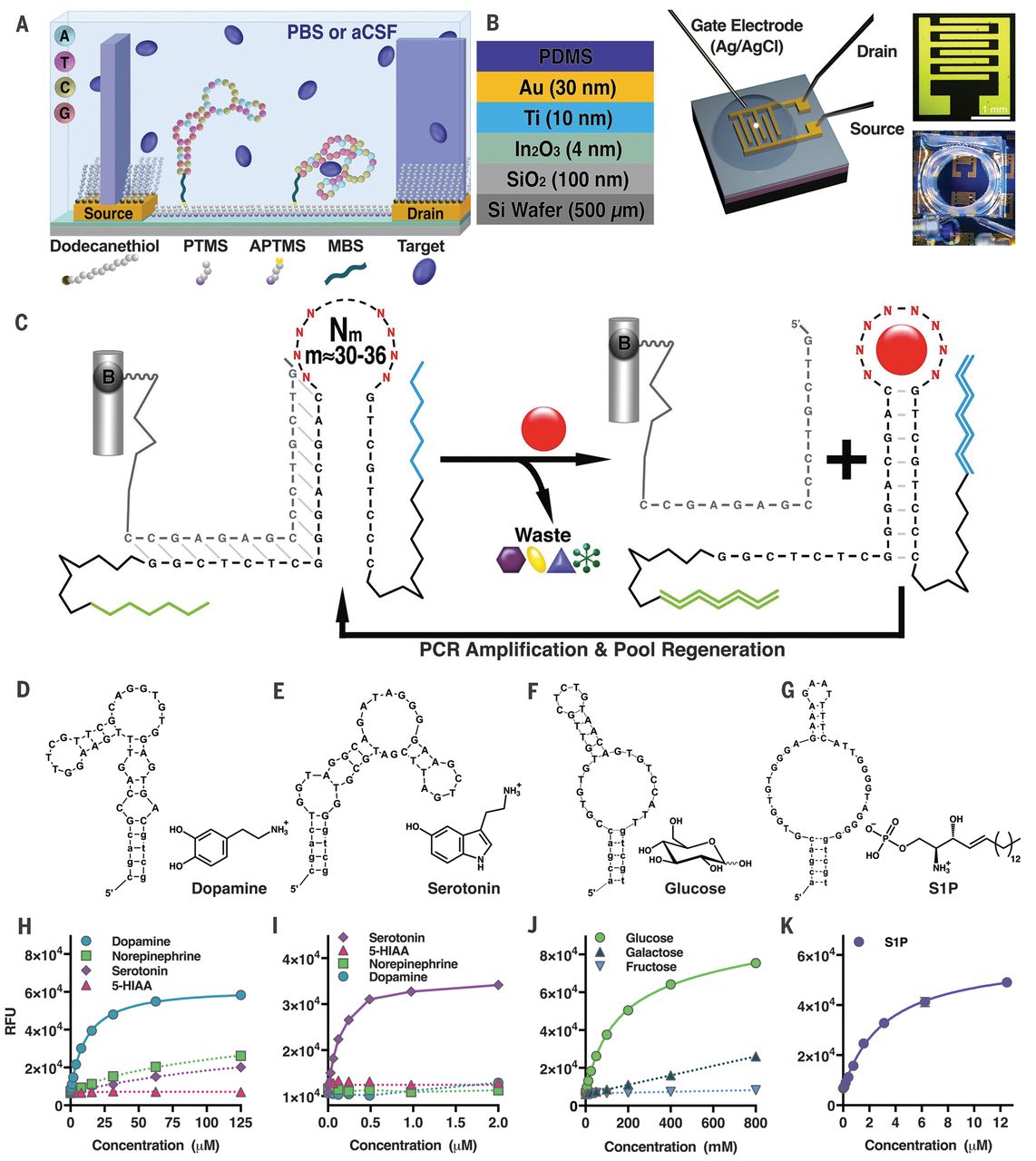
Binding loops stemming from wishful thinking
mFold produces this structure in 1 M NaCl solutions, at 37 °C
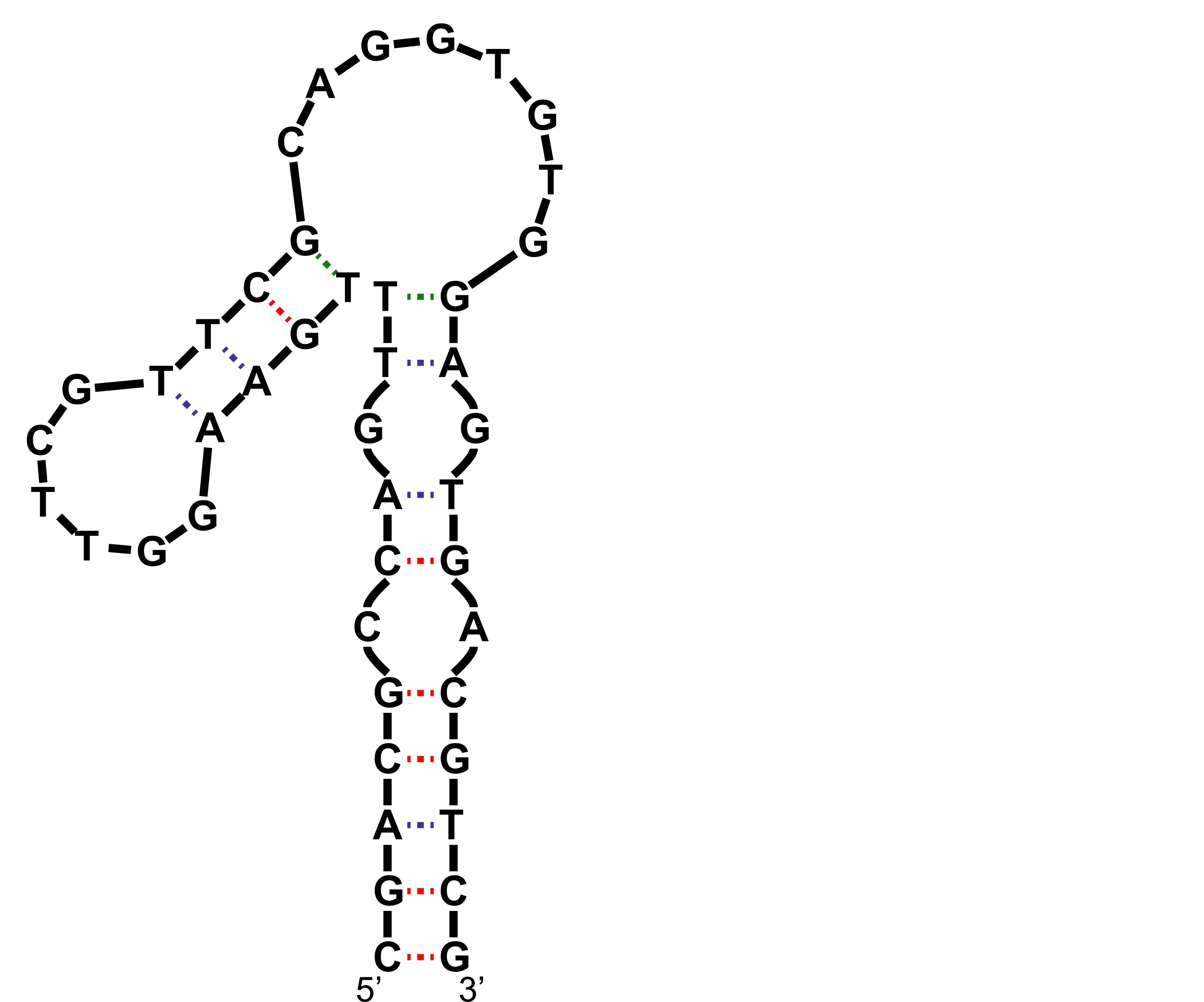
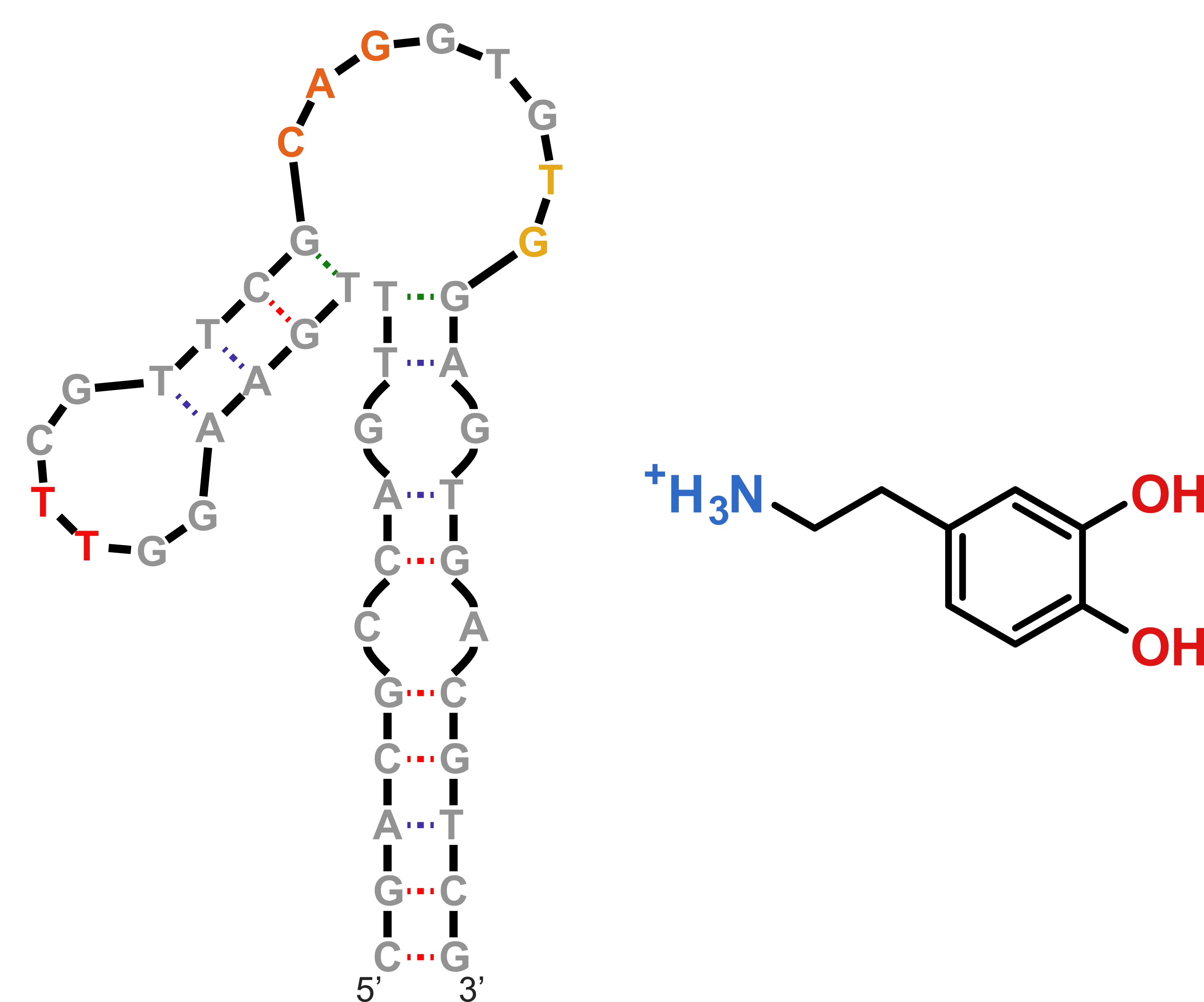
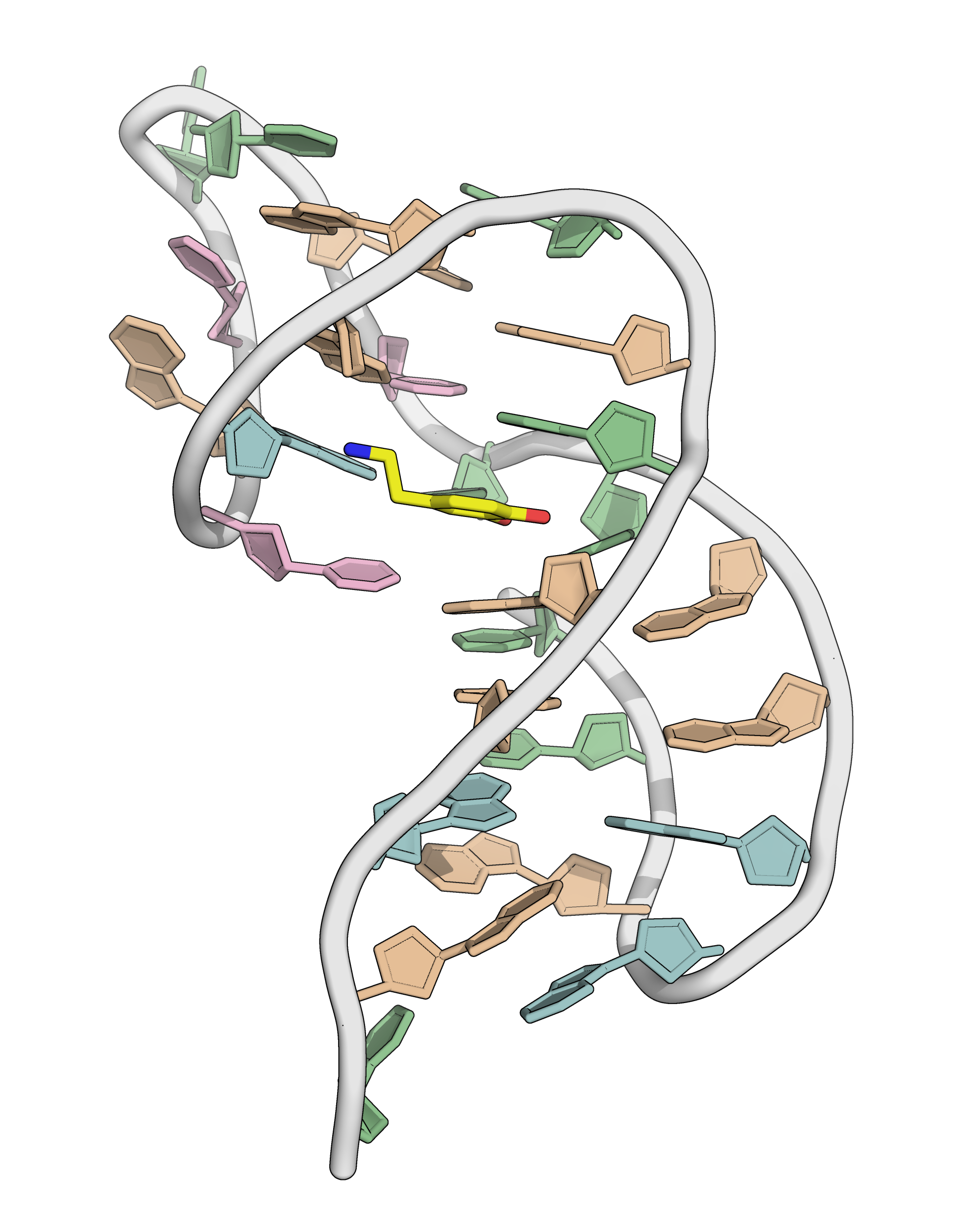
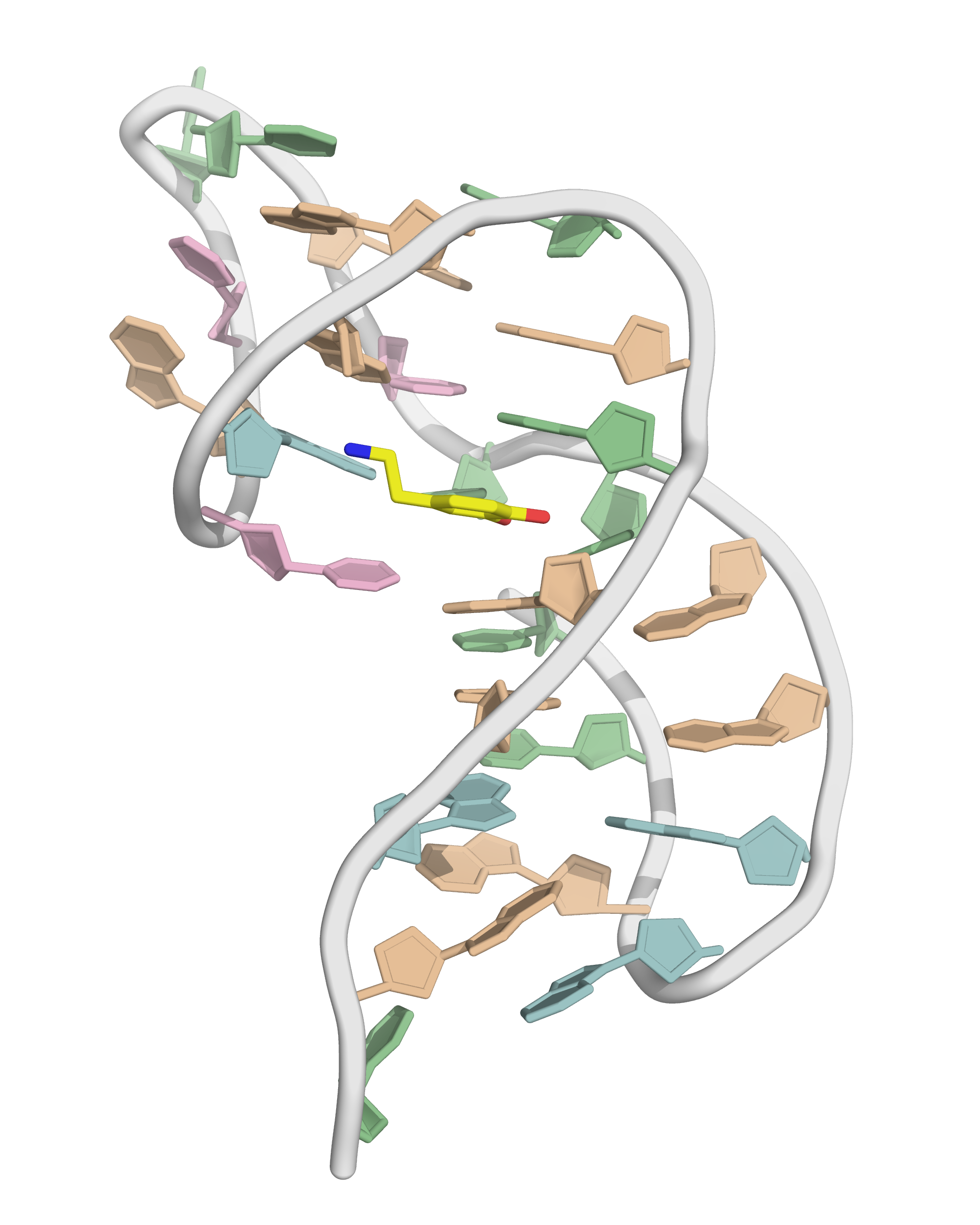
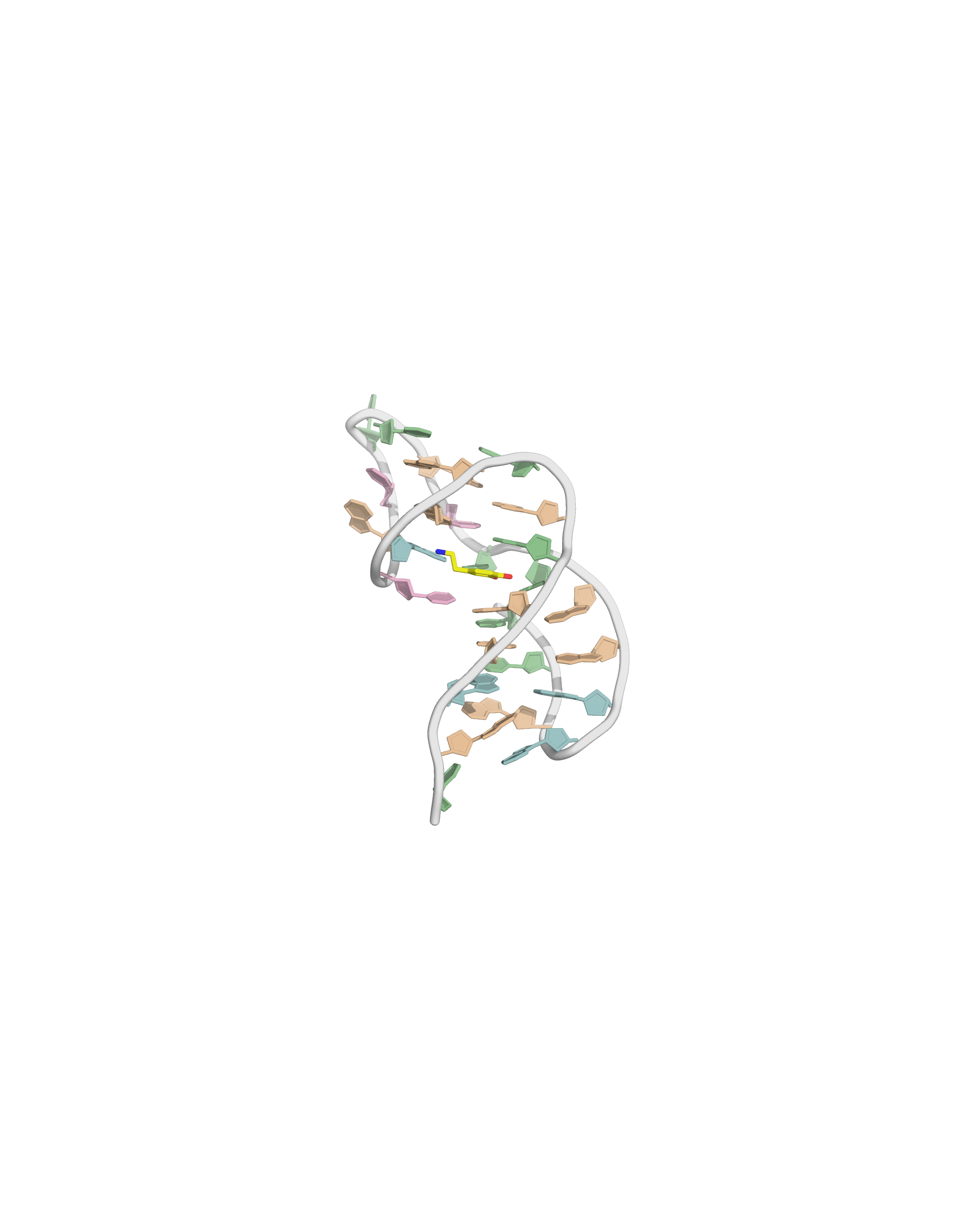
NMR provides a (non-canonical !) starting model
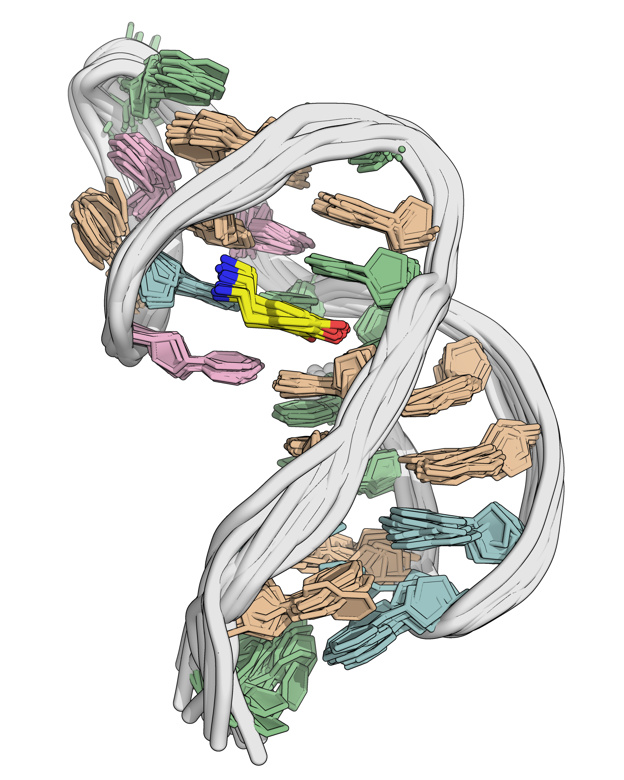
NMR provides an ensemble of models
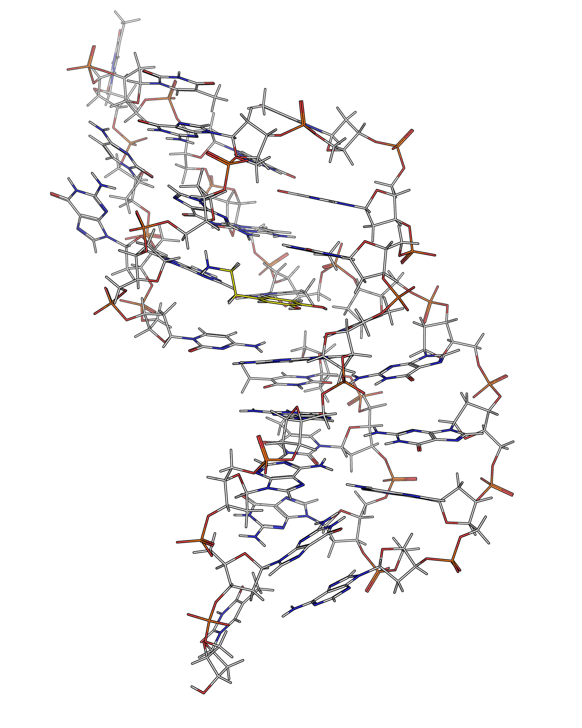
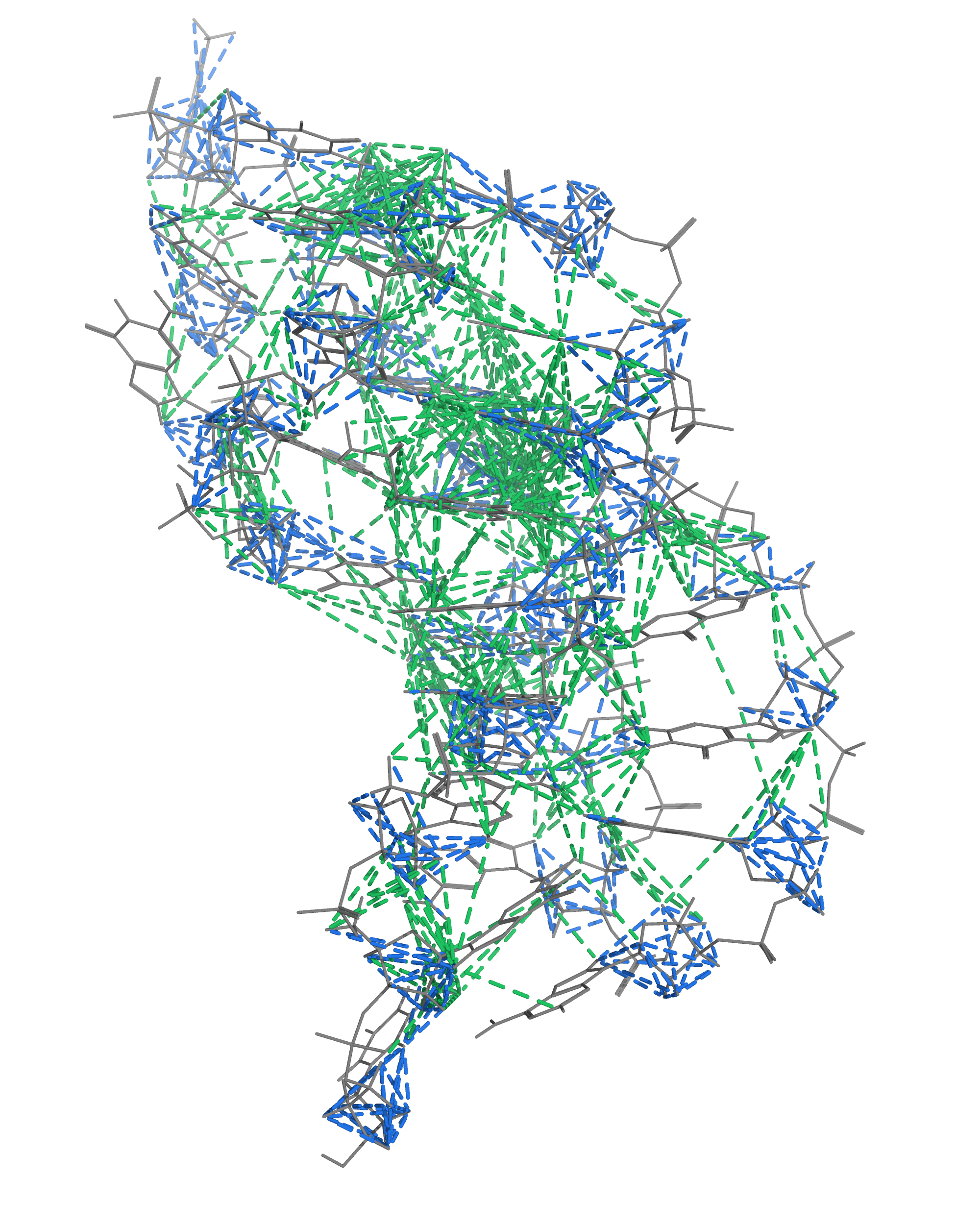
NMR provides many constraints
- DNA (806)
- intra-residue
- inter-residue
- Dopamine (67)
- intramolecular
- intermolecular
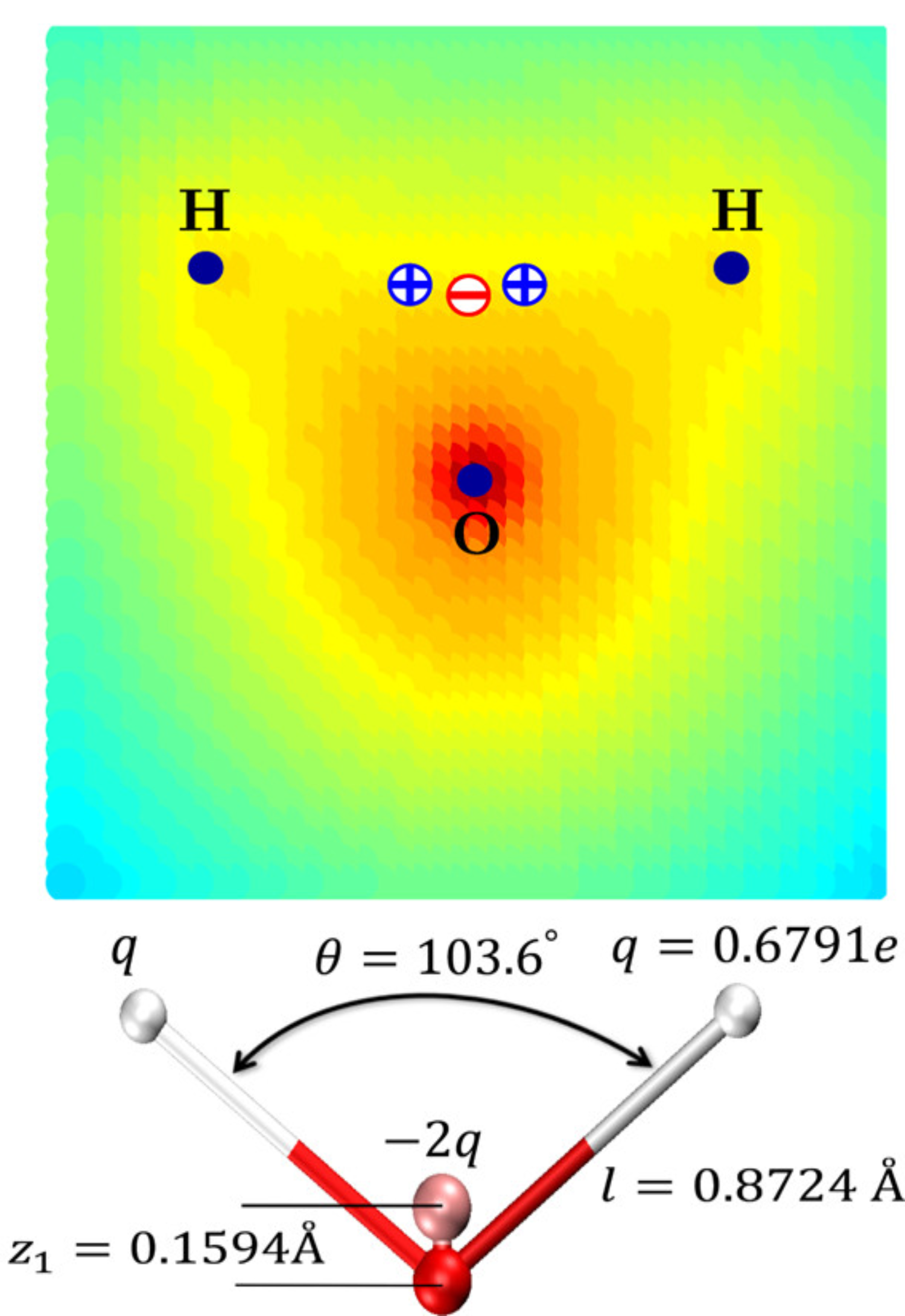
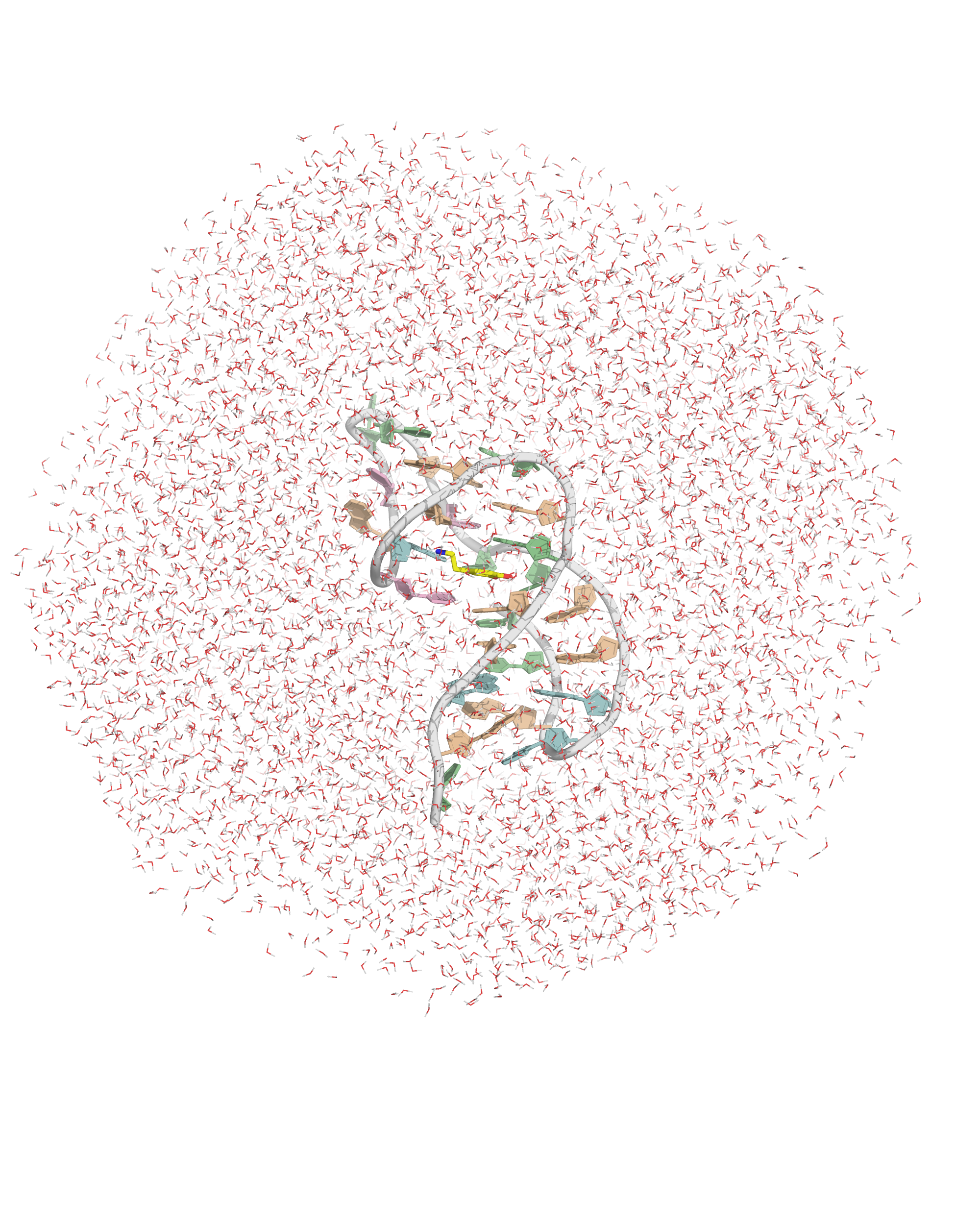
Solvation requires building a box with a model
Solvation requires building a box with a model
- OPC water model
- Octahedral box with, 14 Å to surface
Na+ ions are added to neutralize the system
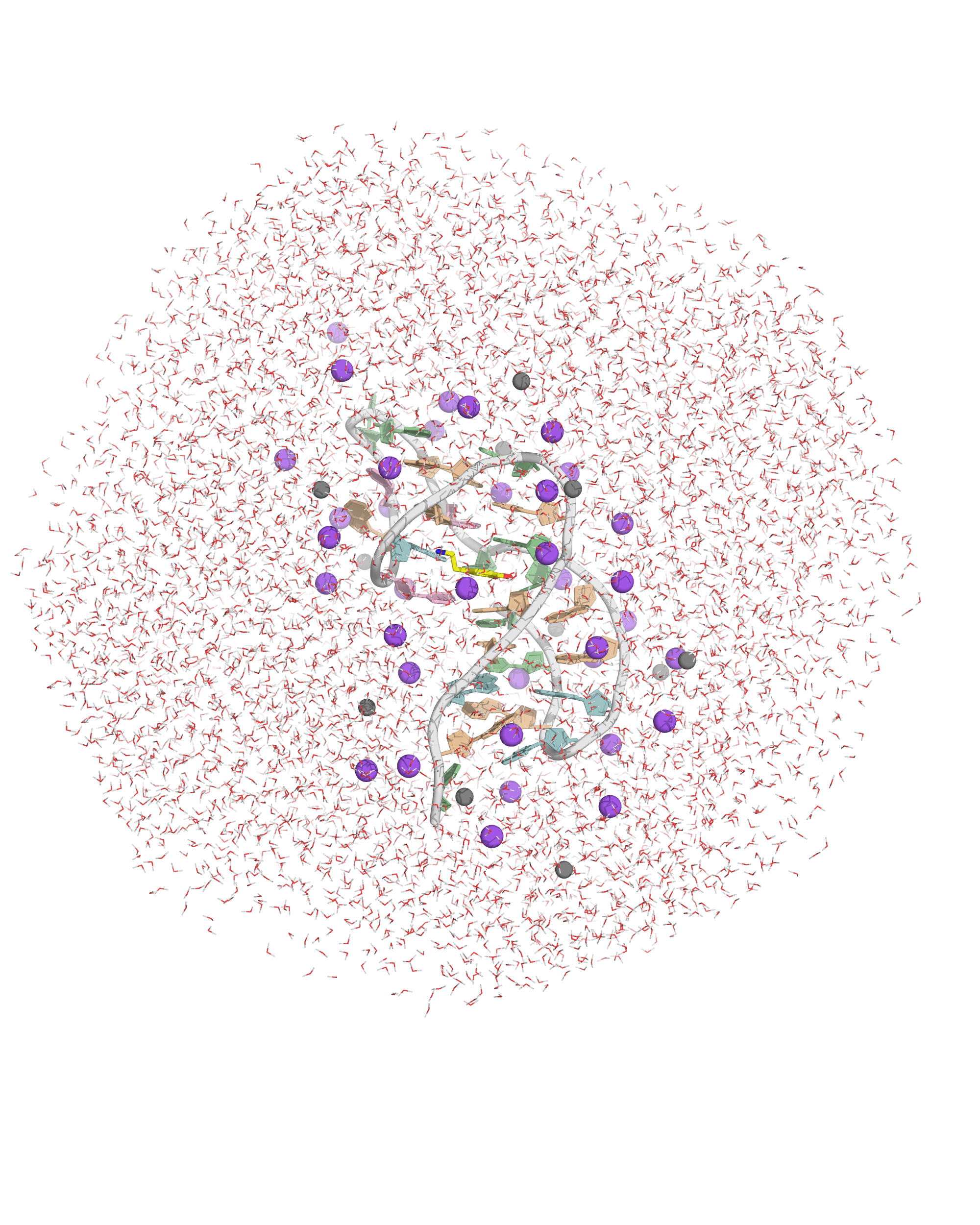
Na+ and Cl- are added to mimic experimental conditions
\(N_{\pm}=\color{#376bd3}{\nu_w}\color{#1a9c1a}{c_0}\color{#000000}e^{\mp \operatorname{arcsinh}\!\left(\frac{\color{#b92424}{Q}}{2\color{#376bd3}{\nu_w}\color{#1a9c1a}{c_0}}\right)}\)
- \(\color{#376bd3}{\nu_w}\): water volume of the box (~ 3x10⁵ Å3)
- \(\color{#1a9c1a}{c_0}\): salt concentration (140 mM)
- \(\color{#b92424}{Q}\): charge (-25)
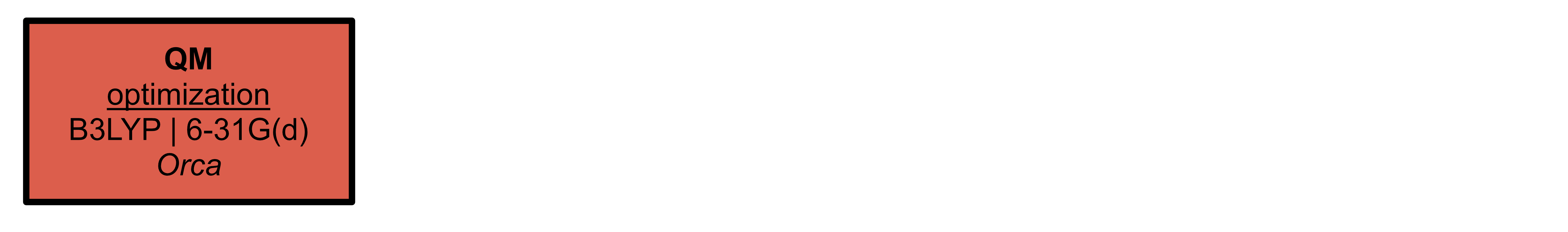
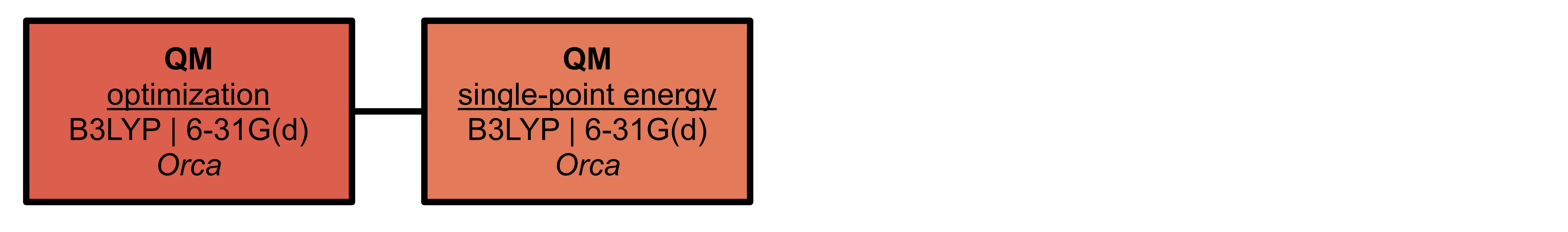
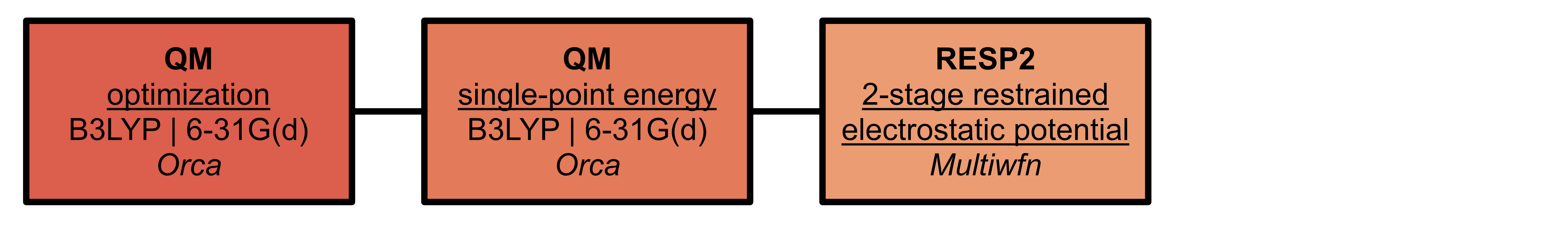
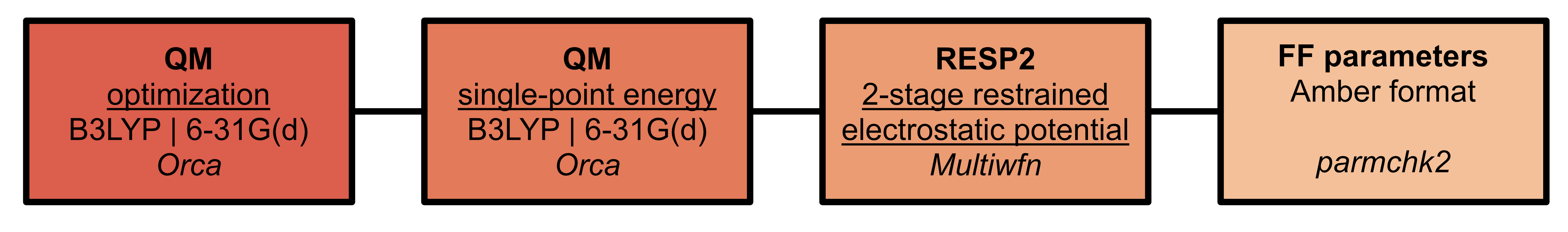
One or several force fields must be used for biomolecules
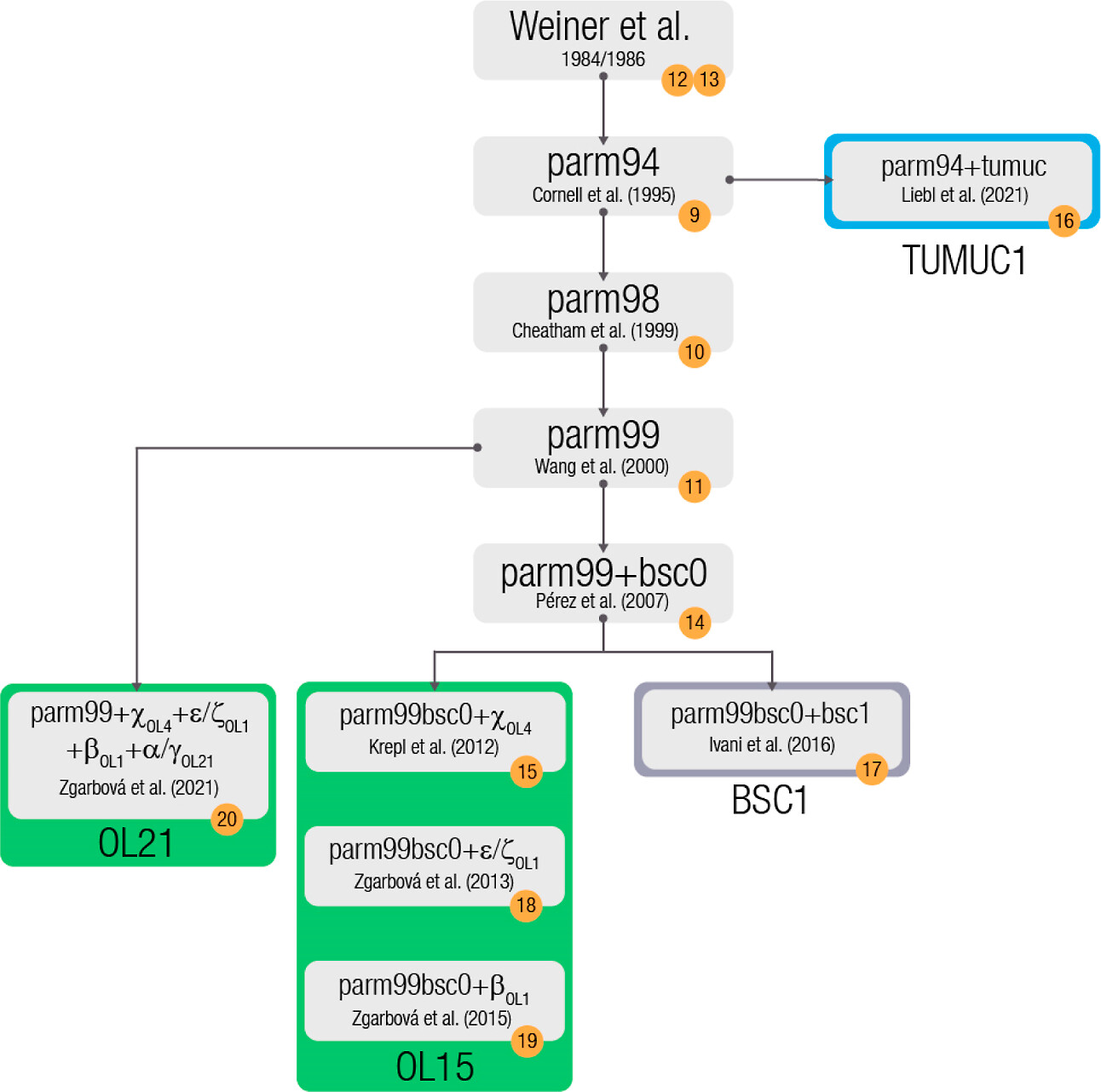
Where necessary, parameters for ligands must be generated
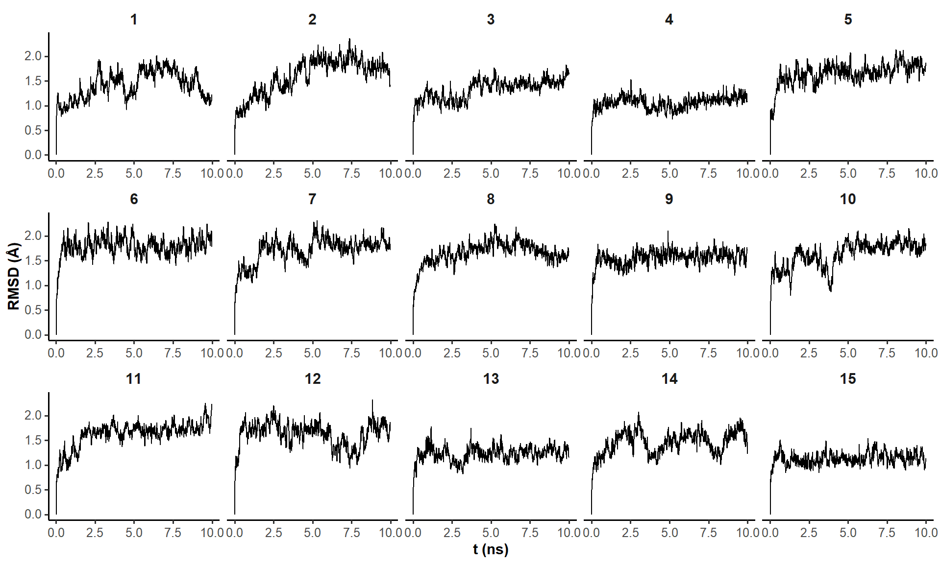
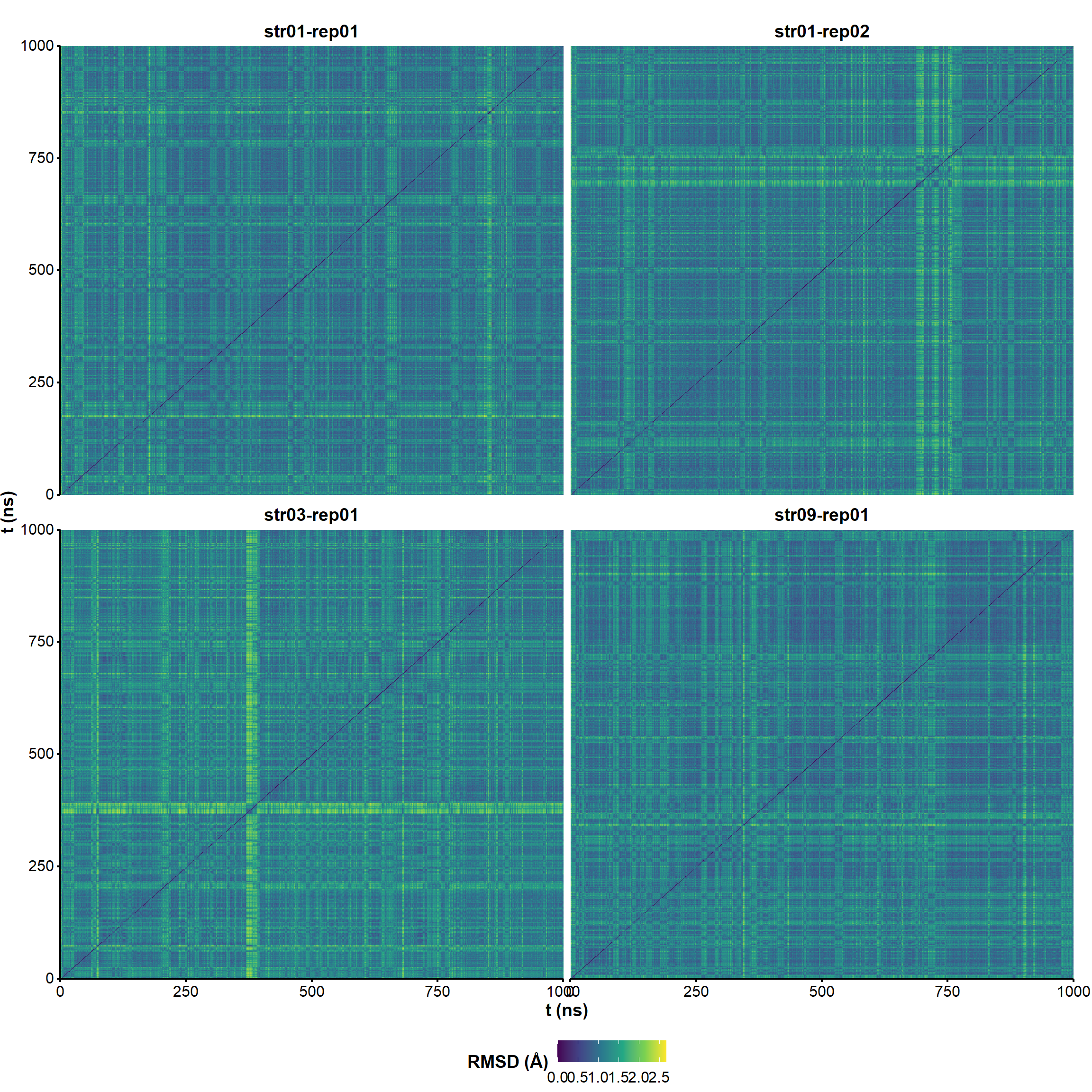
10-ns trajectories are stable RMSD-wise
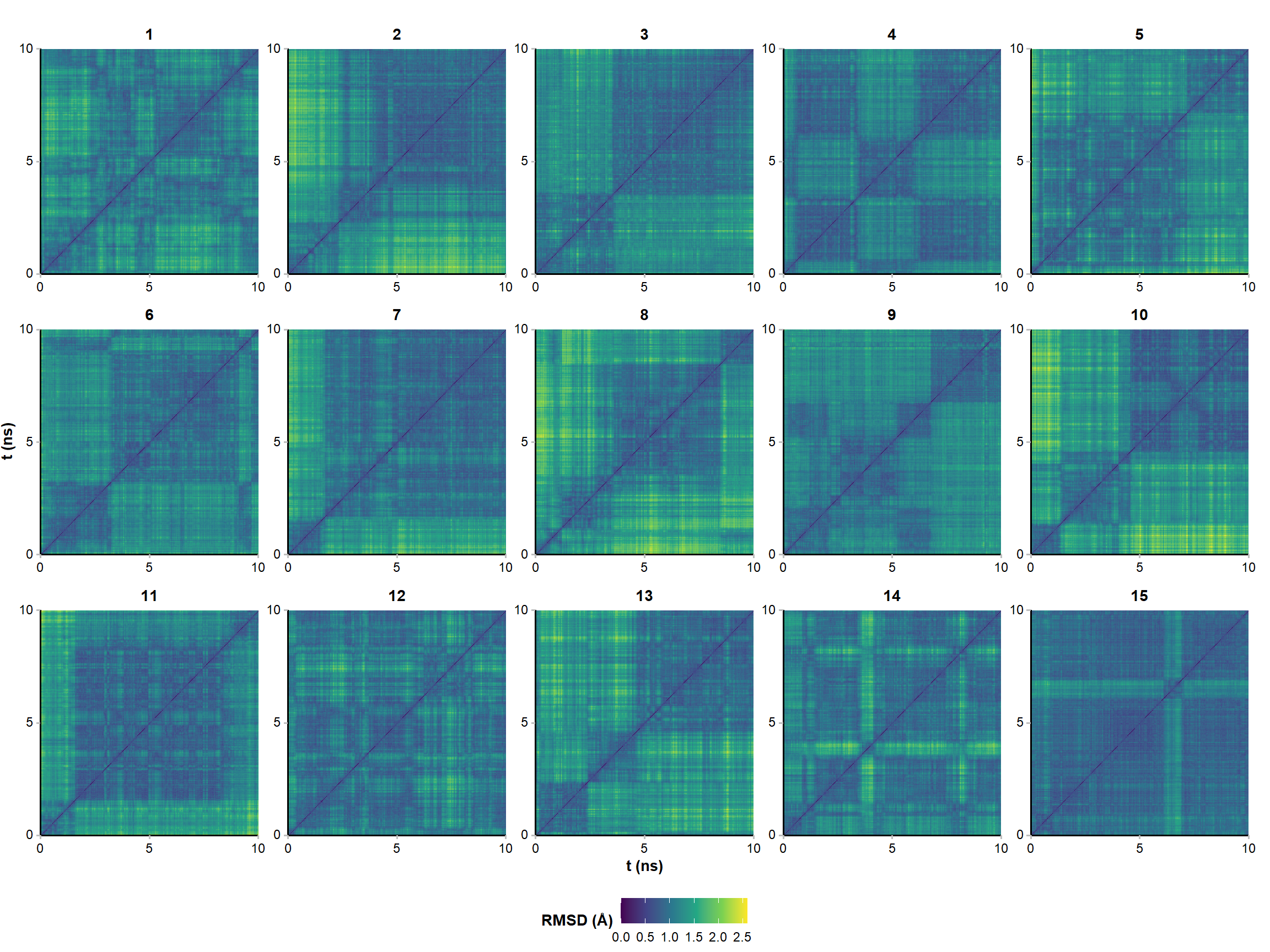
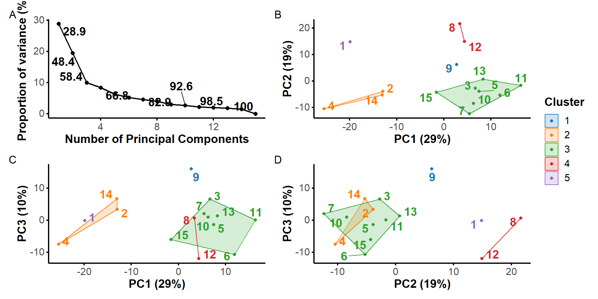
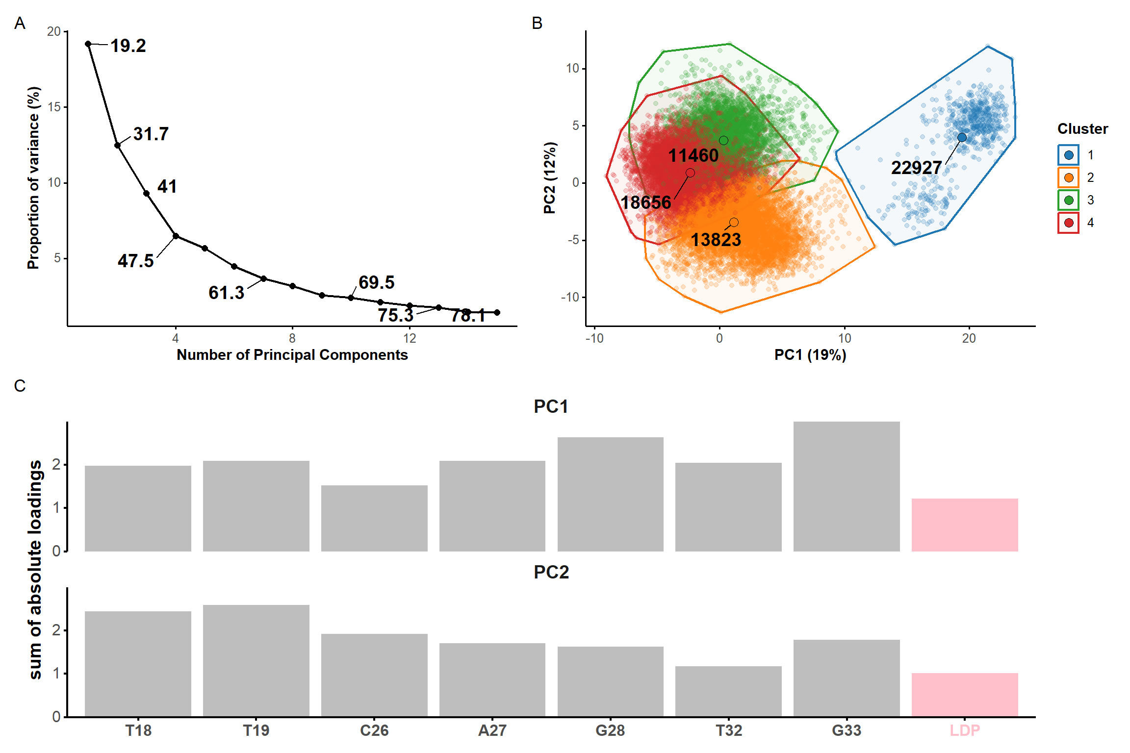
Pairwise RMSD analysis is more informative
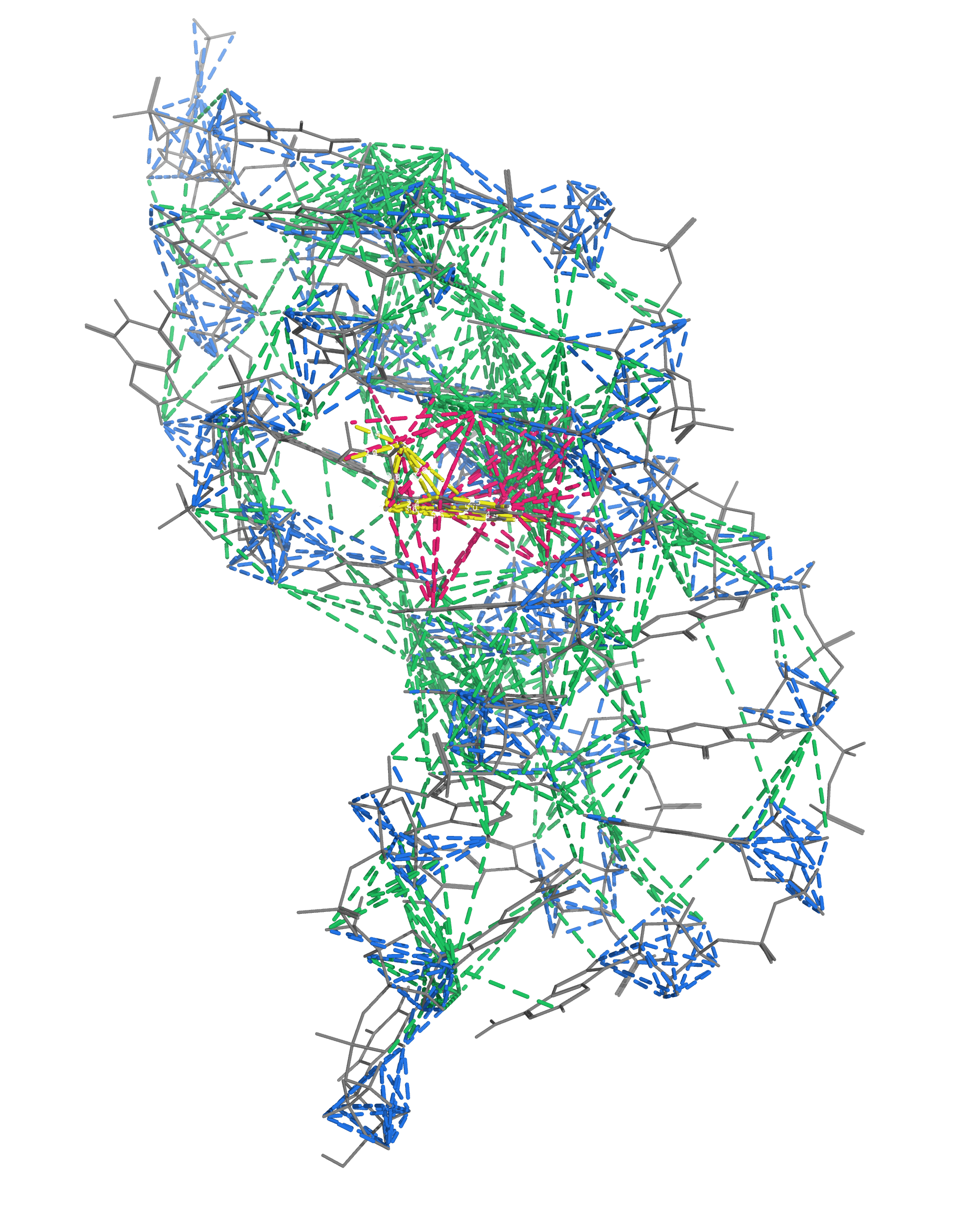
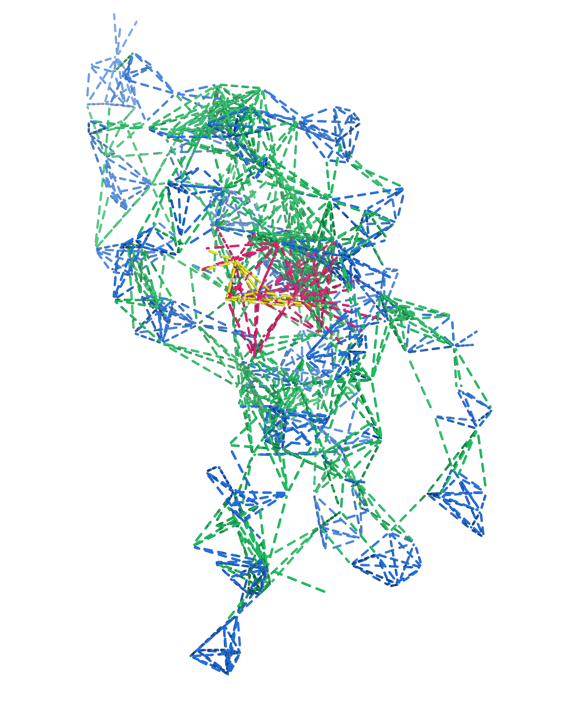
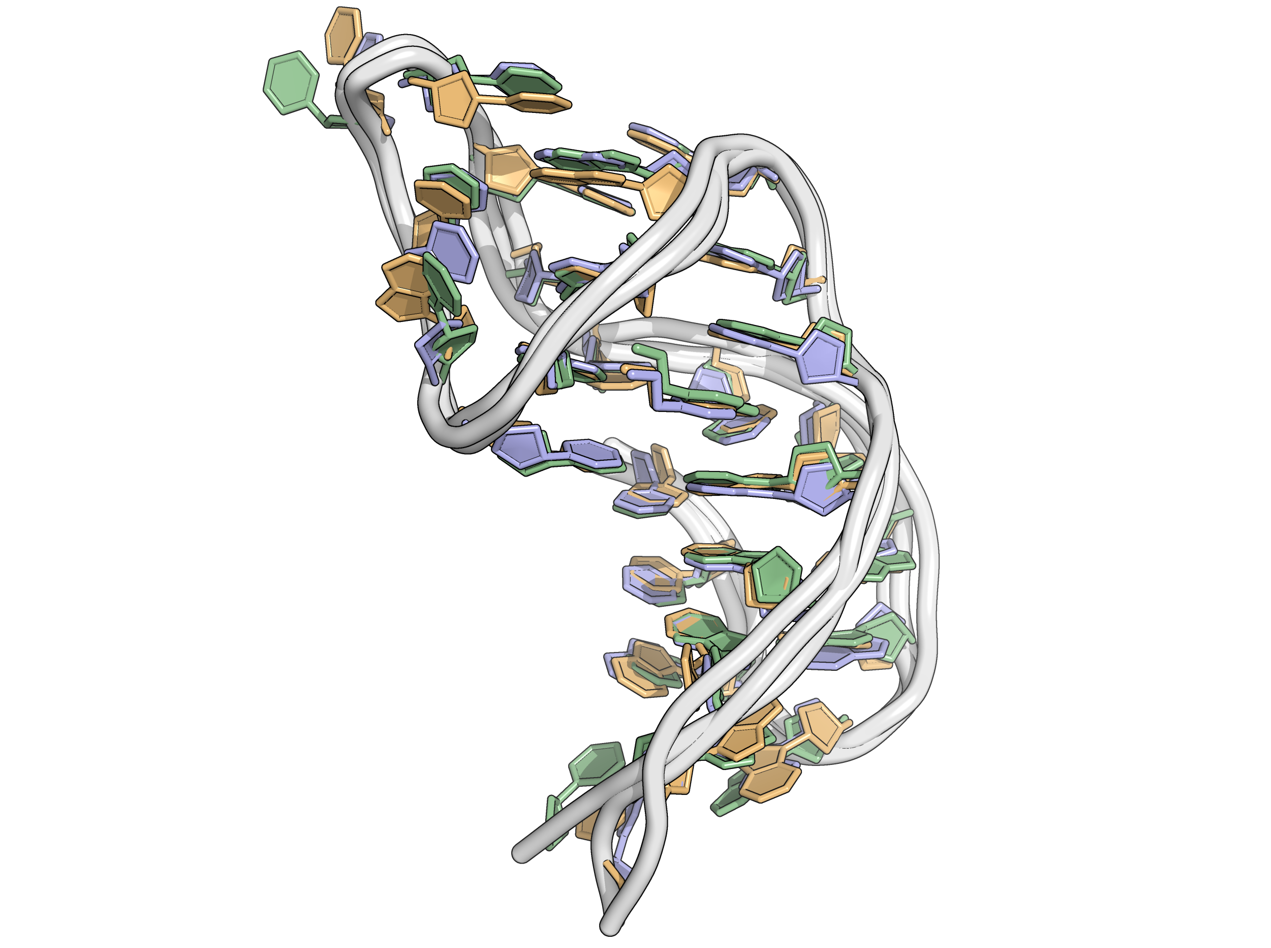
The end point of 15 runs are minimized
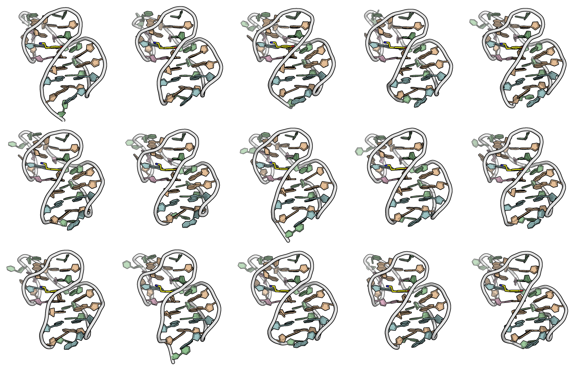
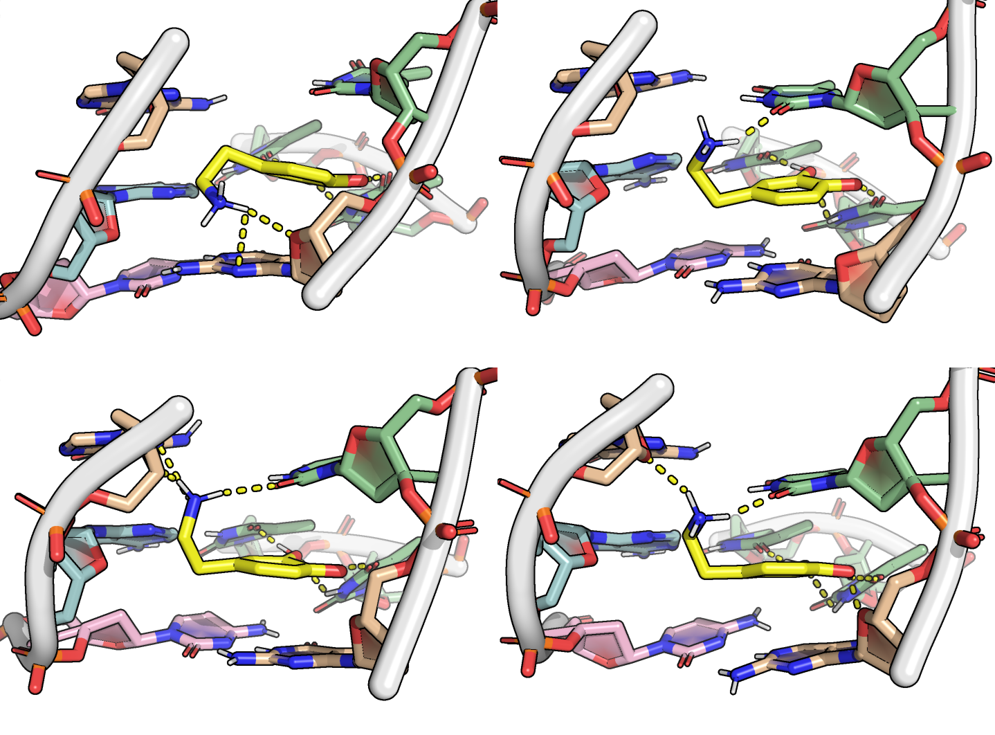
Superimposition reveals a well-defined binding pocket
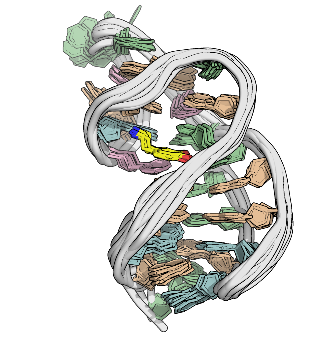
NMR restraints are satisfied
Replication is necessary (at least to publish!)
Longer simulations are stable and reproducible
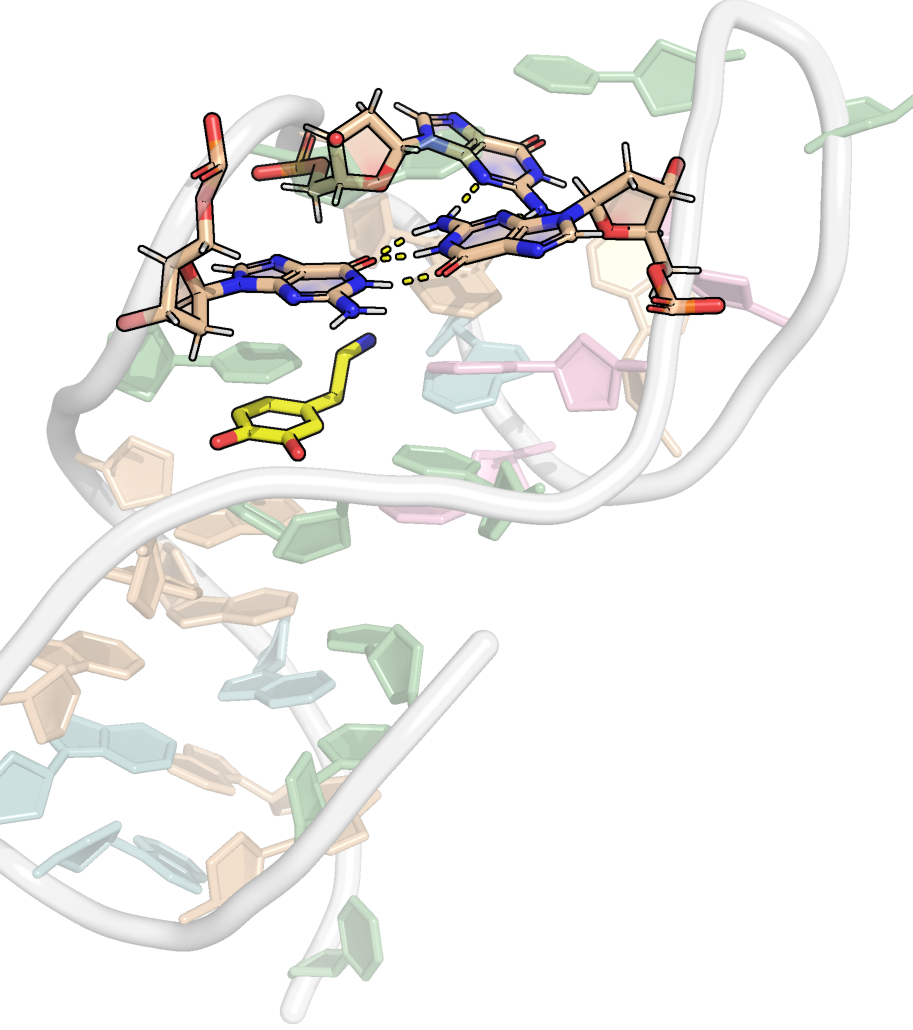
Non-WC base pairs and triplets
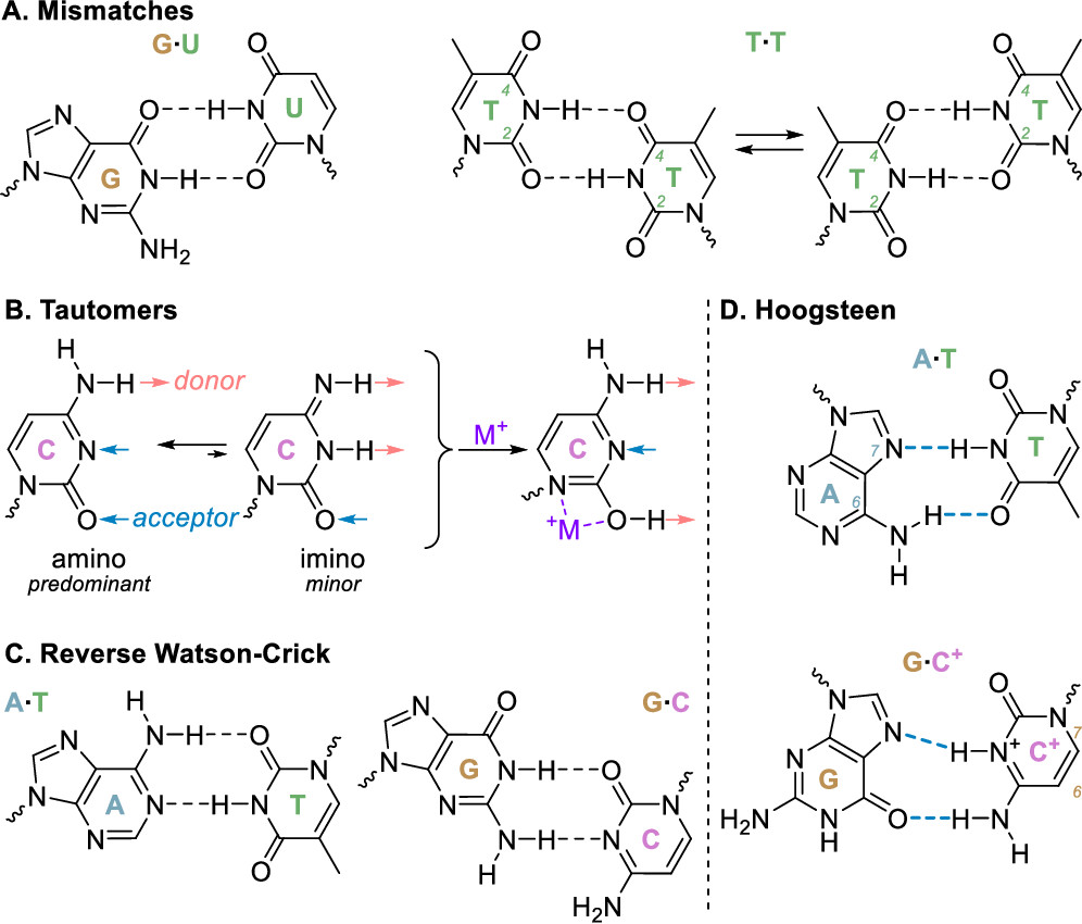
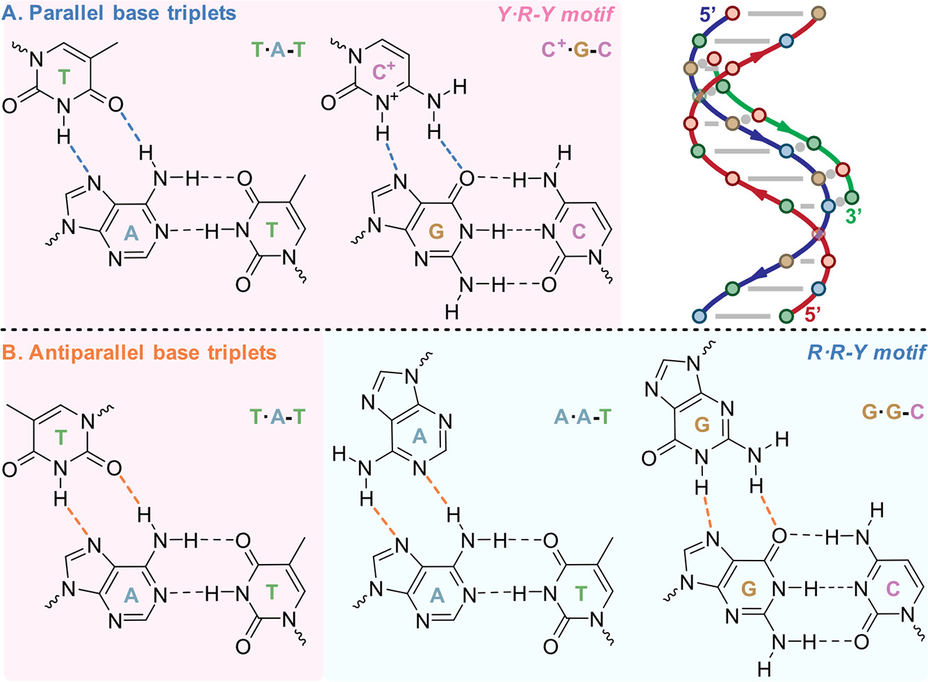

Only two WC base pairs are present
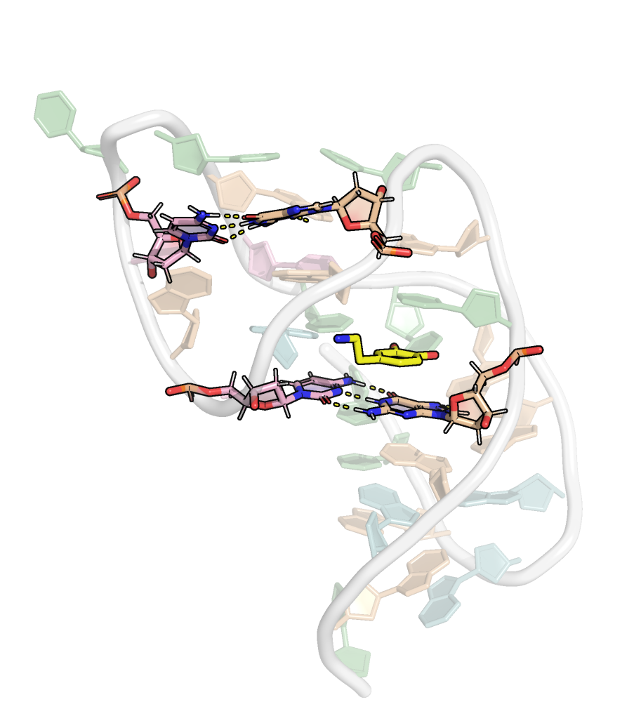
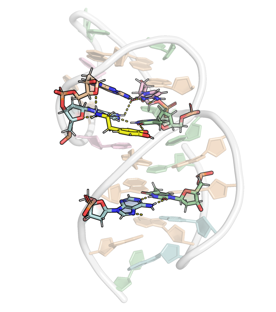
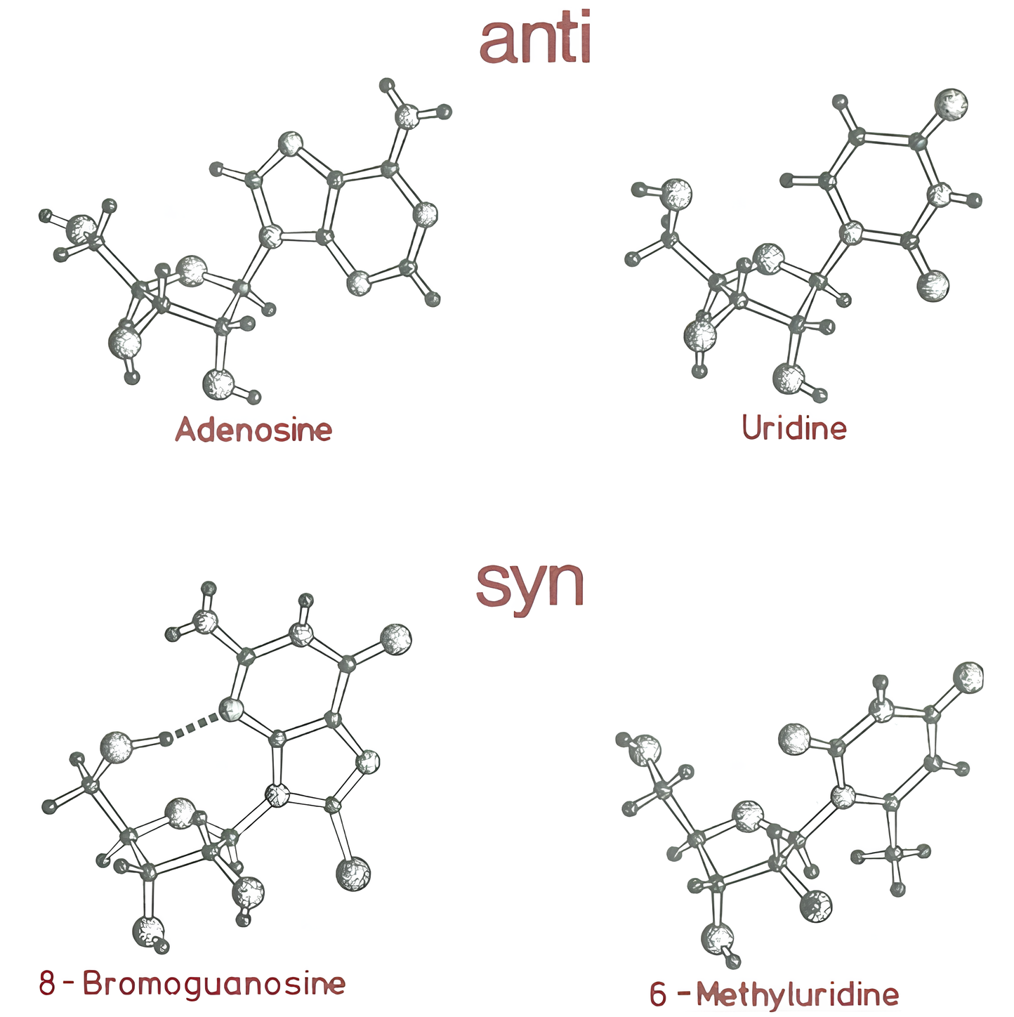
Presence of base triplets with syn glycosidic bond angle
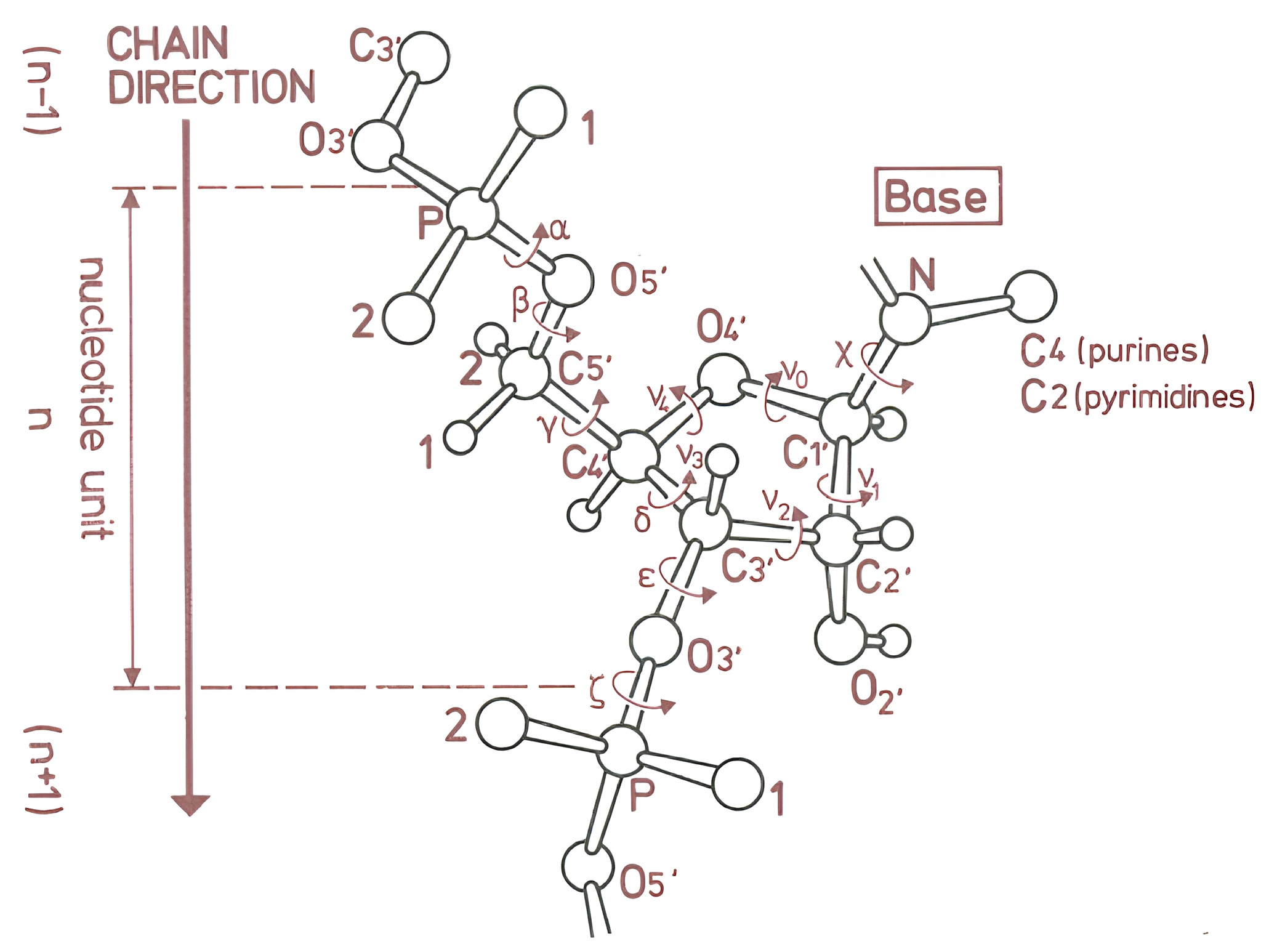
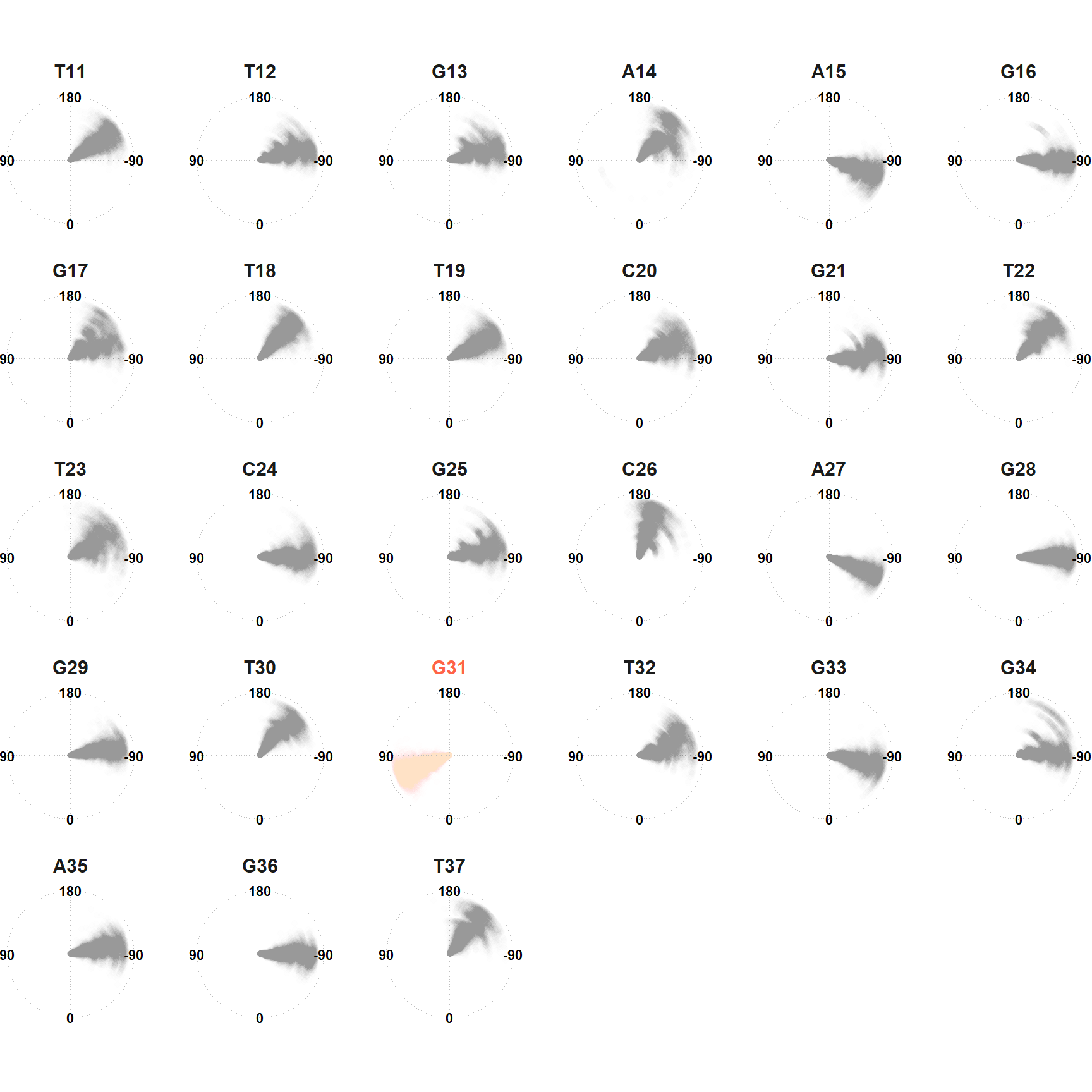
Dihedral angles are stable along the simulation

Binding requires non-canonical puckers
\[\color{#F0A308}\theta_M \color{#000000}= \frac{\color{#376bd3}\nu_2}{\cos \color{#279400}P}\]
\[\tan \color{#279400}P \color{#000000}= \frac{(\color{#008594}\nu_4 \color{#000000}+ \color{#A91FB6}\nu_1\color{#000000})-(\color{#940000}\nu_3 \color{#000000}+ \color{#947E00}\nu_0\color{#000000})}{2\color{#376bd3}\nu_2\color{#000000}(\sin(\frac{\pi}{5}) + \sin(\frac{2\pi}{5}))}\]
Binding requires non-canonical puckers
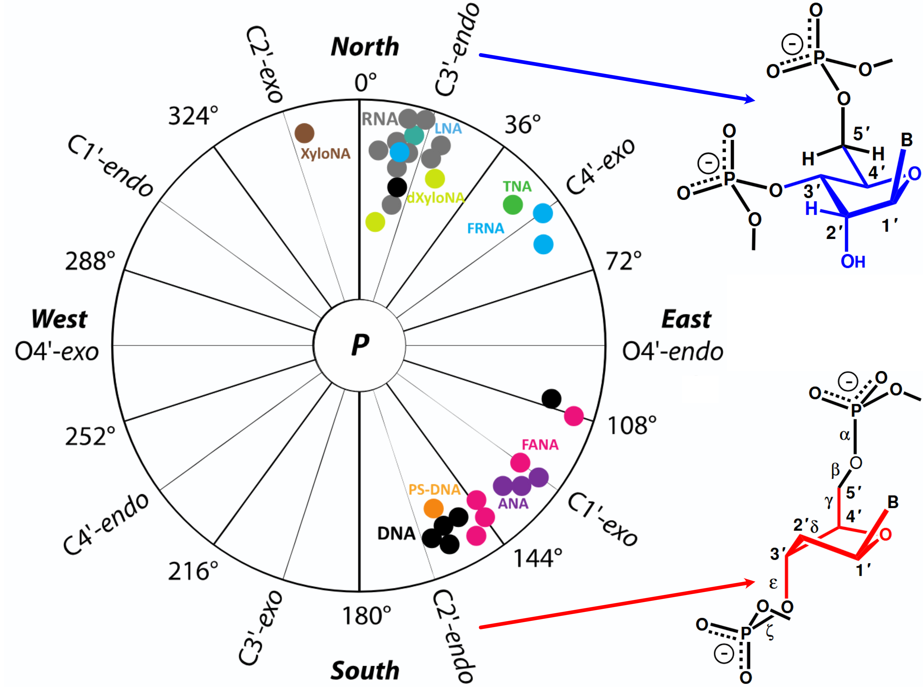
Binding requires non-canonical puckers
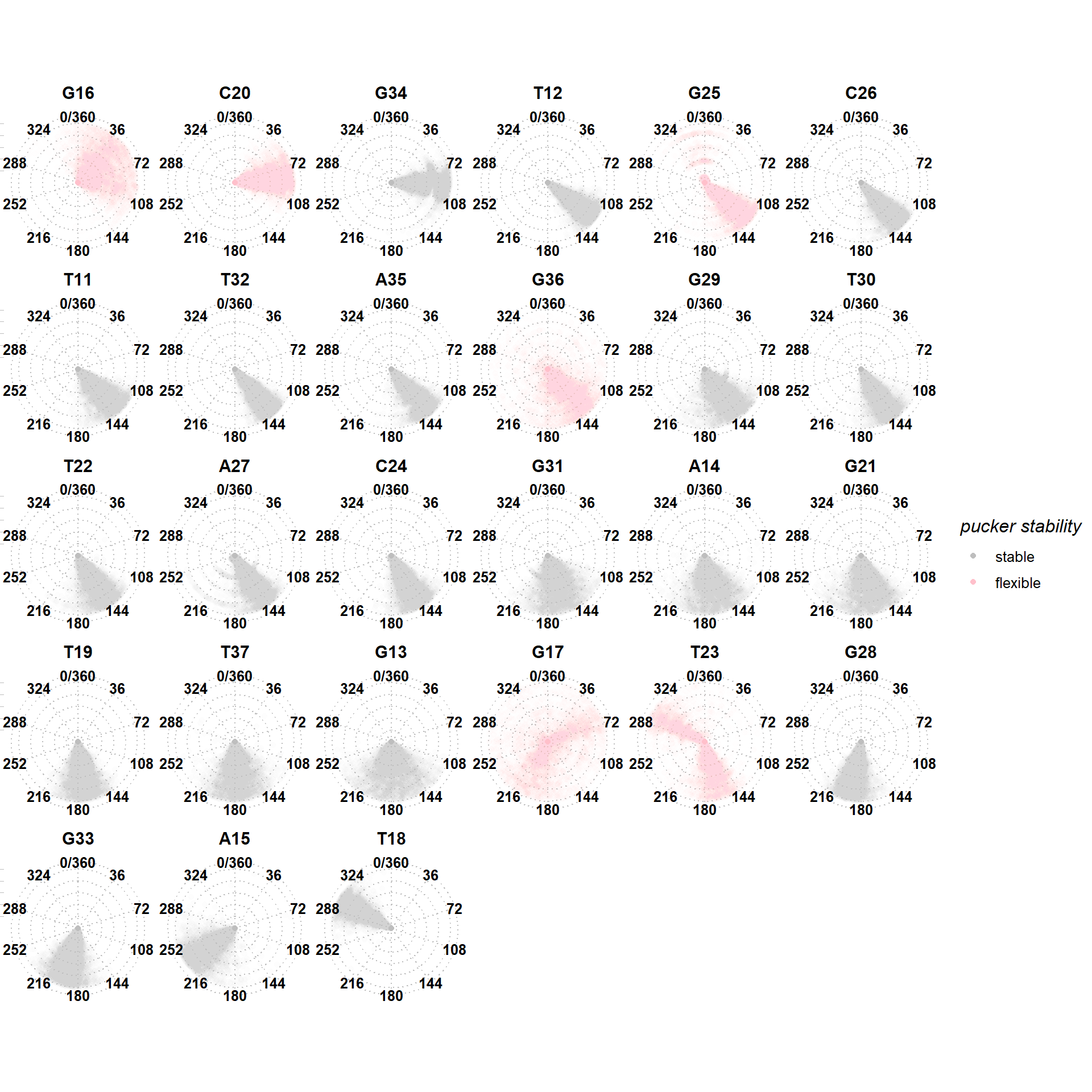
Binding requires non-canonical puckers
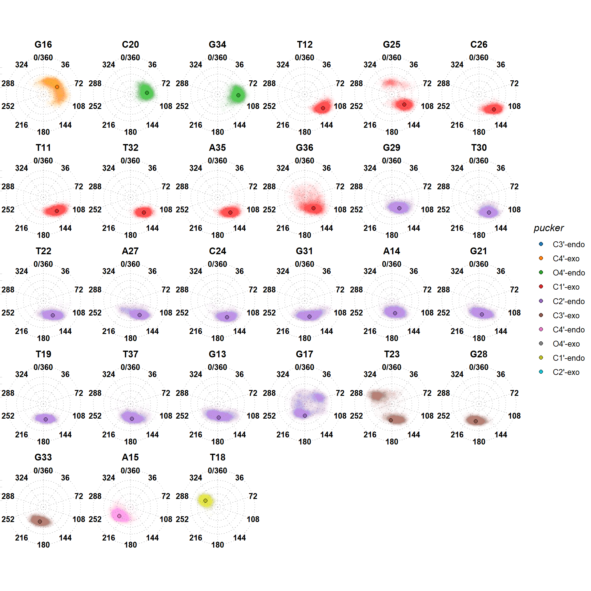
T18 adopts a C1’-endo pucker upon binding
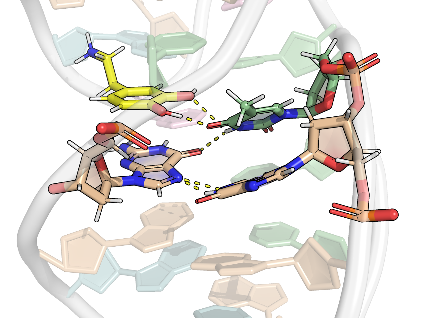
Binding through H-bonds, electrostatic and π interactions
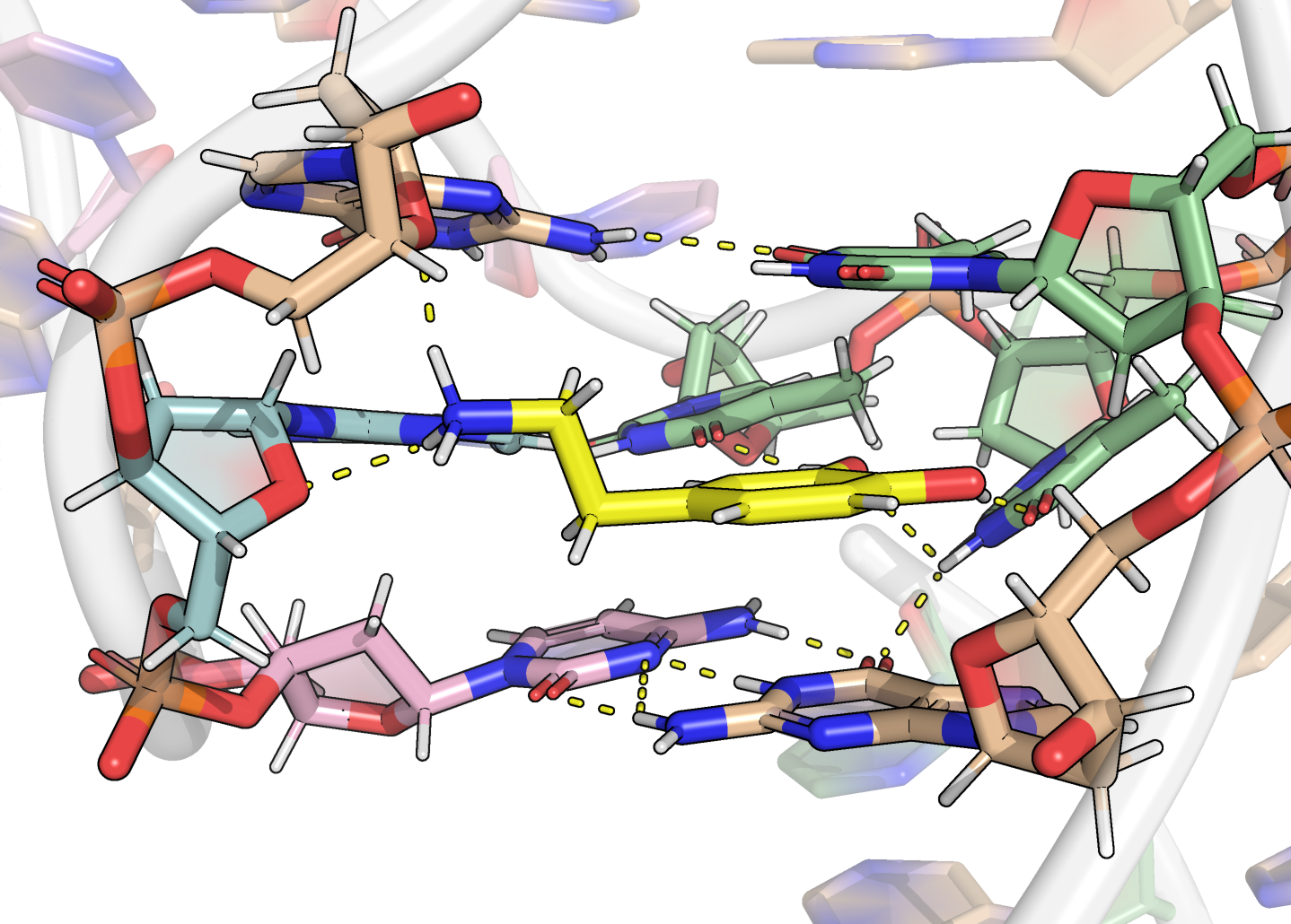
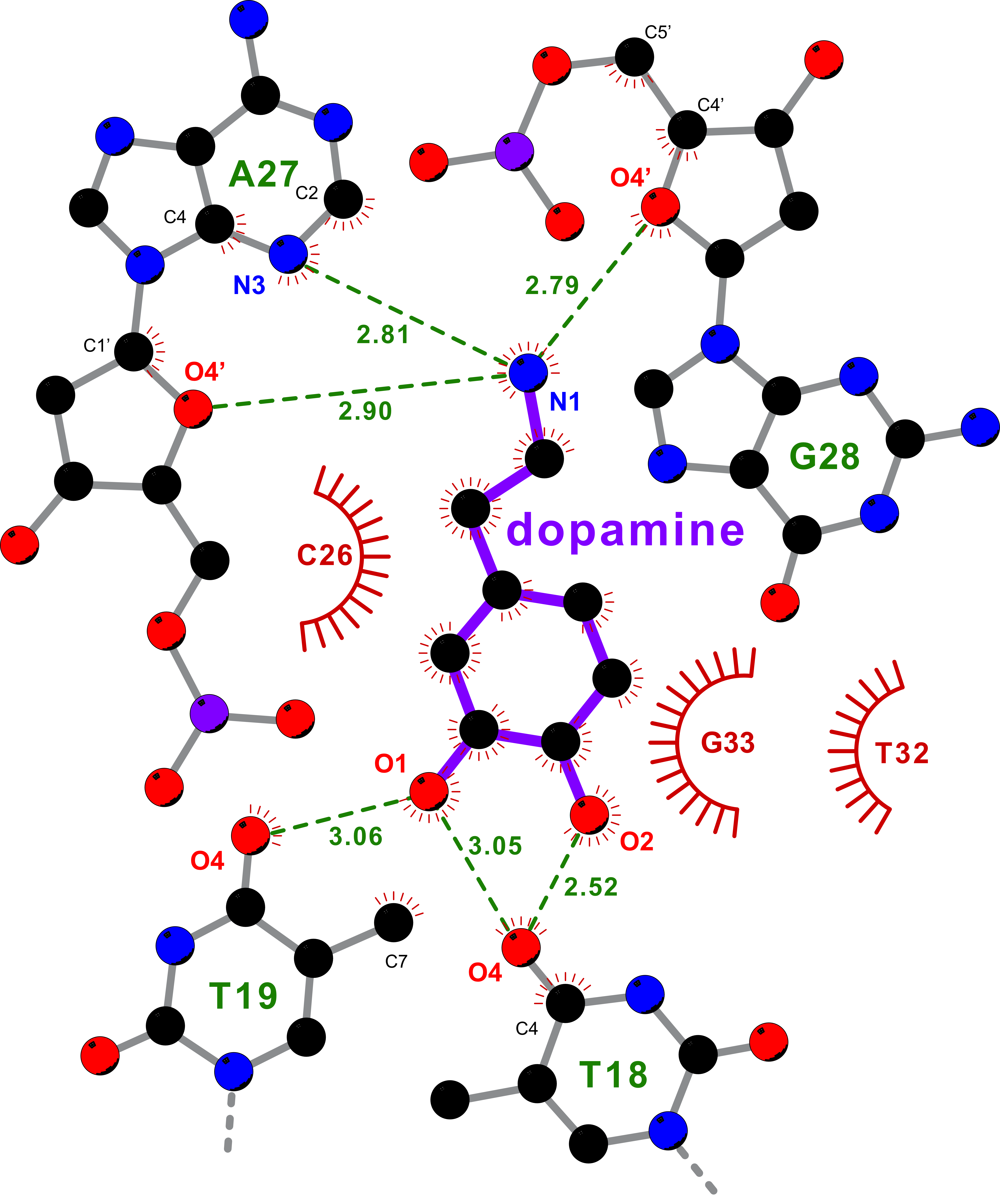

Restraints are necessary to refine the binding mode
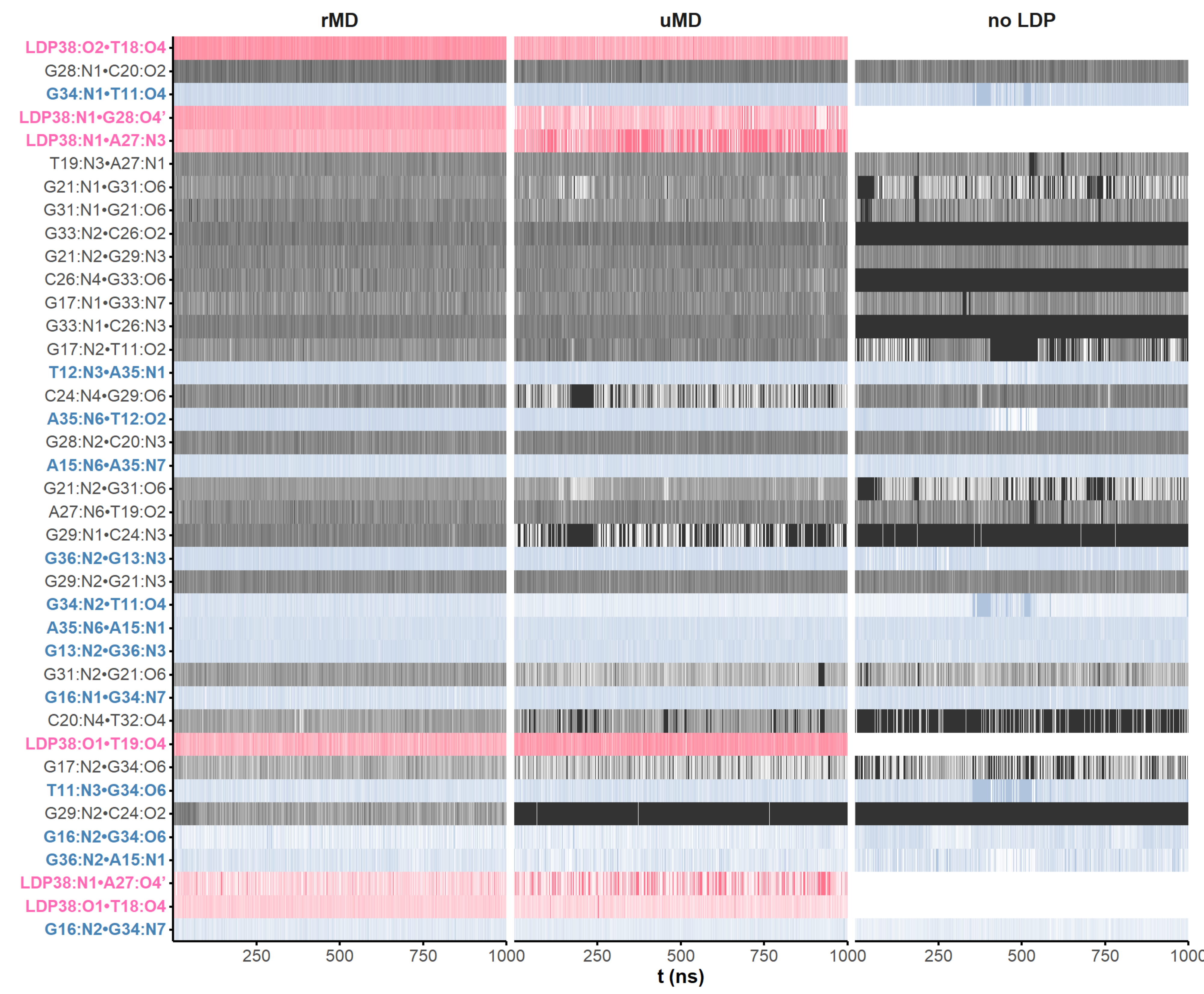
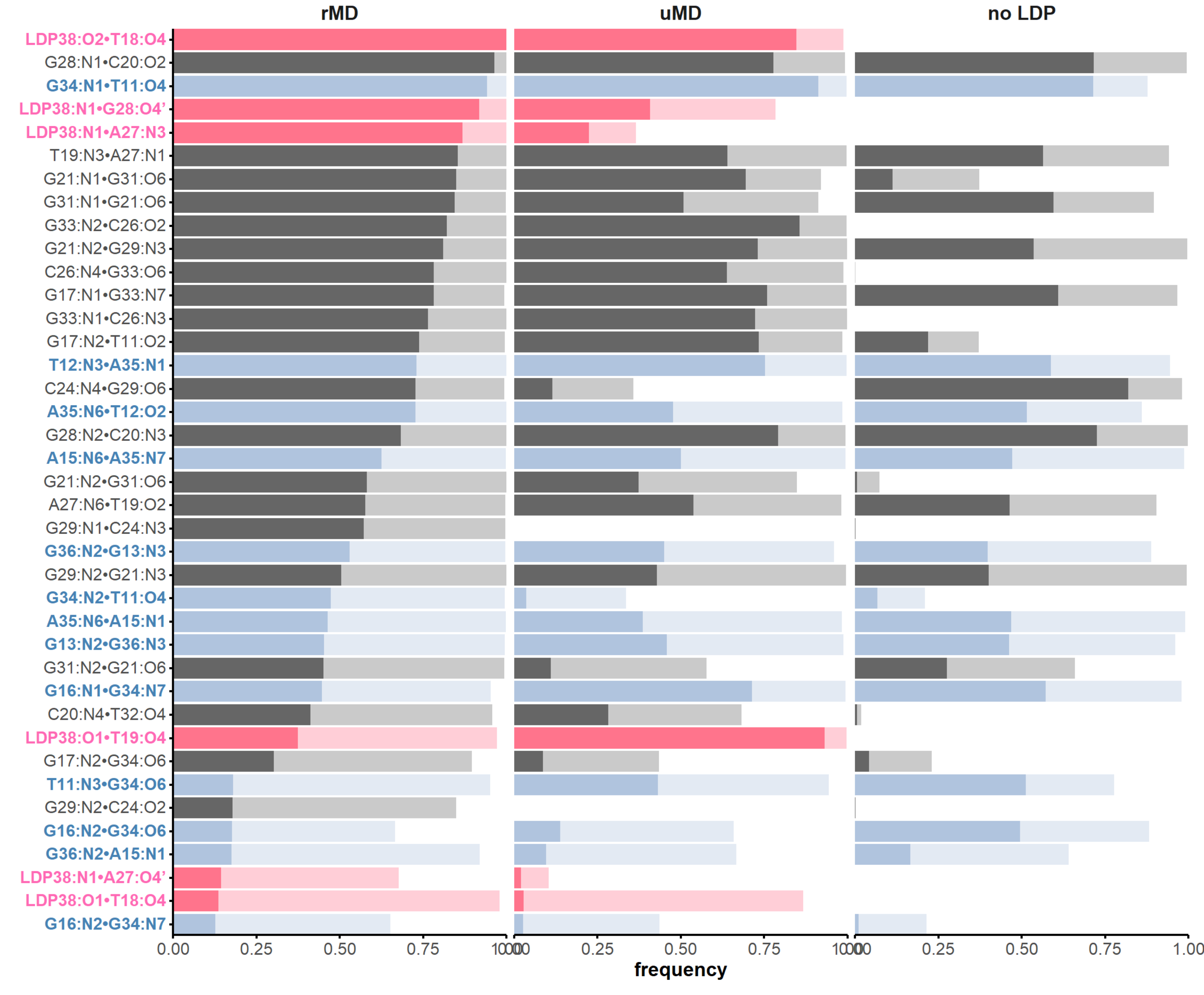
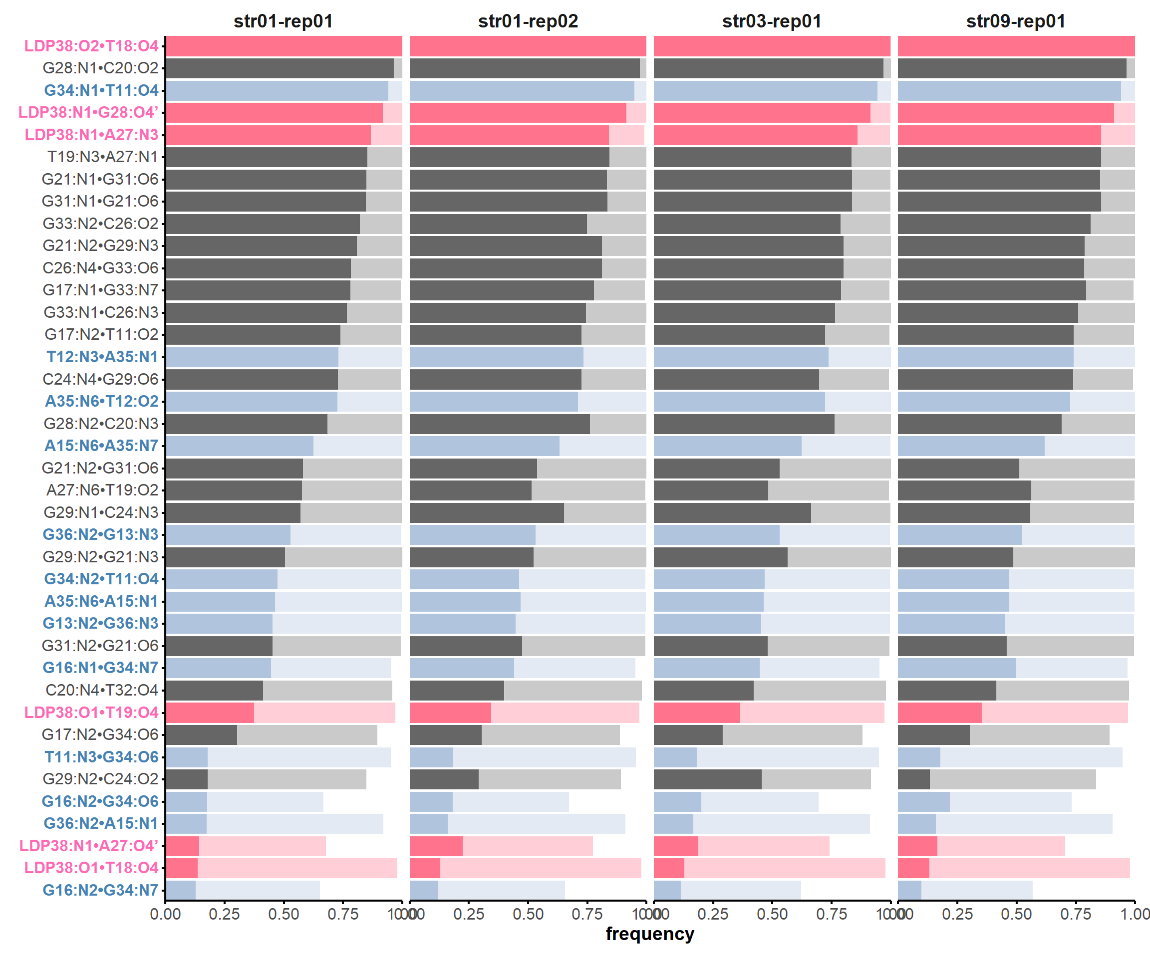
H-bonds inform structural dynamics
and model robustness
Prediction models still come short for aptamers
- 0 out of 107 submitted models with \(RMSD < 10 Å\)
- Training set is not diverse enough
- Dopamine essential for folding
